


 Foundational characteristics of cancer include proliferation, angiogenesis, migration, evasion of apoptosis, and cellular immortality. Find key markers for these cellular processes and antibodies to detect them.
 The SUMOplot™ Analysis Program predicts and scores sumoylation sites in your protein. SUMOylation is a post-translational modification involved in various cellular processes, such as nuclear-cytosolic transport, transcriptional regulation, apoptosis, protein stability, response to stress, and progression through the cell cycle.
 The Autophagy Receptor Motif Plotter predicts and scores autophagy receptor binding sites in your protein. Identifying proteins connected to this pathway is critical to understanding the role of autophagy in physiological as well as pathological processes such as development, differentiation, neurodegenerative diseases, stress, infection, and cancer.
MALT1 Polyclonal Antibody
- SPECIFICATION
- CITATIONS
- PROTOCOLS
- BACKGROUND

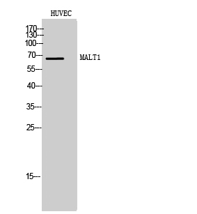
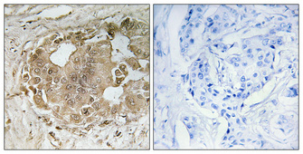
Application
| WB, IHC-P, IF |
|---|---|
| Primary Accession | Q9UDY8 |
| Reactivity | Human, Mouse |
| Host | Rabbit |
| Clonality | Polyclonal |
| Calculated MW | 92272 Da |
| Gene ID | 10892 |
|---|---|
| Other Names | MALT1; MLT; Mucosa-associated lymphoid tissue lymphoma translocation protein 1; MALT lymphoma-associated translocation; Paracaspase |
| Dilution | WB~~Western Blot: 1/500 - 1/2000. Immunohistochemistry: 1/100 - 1/300. Immunofluorescence: 1/200 - 1/1000. ELISA: 1/10000. Not yet tested in other applications. IHC-P~~N/A IF~~1:50~200 |
| Format | Liquid in PBS containing 50% glycerol, 0.5% BSA and 0.09% (W/V) sodium azide. |
| Storage Conditions | -20℃ |
| Name | MALT1 {ECO:0000303|PubMed:10523859, ECO:0000312|HGNC:HGNC:6819} |
|---|---|
| Function | Protease that enhances BCL10-induced activation: acts via formation of CBM complexes that channel adaptive and innate immune signaling downstream of CARD domain-containing proteins (CARD9, CARD11 and CARD14) to activate NF-kappa-B and MAP kinase p38 pathways which stimulate expression of genes encoding pro-inflammatory cytokines and chemokines (PubMed:11262391, PubMed:18264101, PubMed:24074955). Mediates BCL10 cleavage: MALT1-dependent BCL10 cleavage plays an important role in T-cell antigen receptor-induced integrin adhesion (PubMed:11262391, PubMed:18264101). Involved in the induction of T helper 17 cells (Th17) differentiation (PubMed:11262391, PubMed:18264101). Cleaves RC3H1 and ZC3H12A in response to T-cell receptor (TCR) stimulation which releases their cooperatively repressed targets to promote Th17 cell differentiation (By similarity). Also mediates cleavage of N4BP1 in T-cells following TCR-mediated activation, leading to N4BP1 inactivation (PubMed:31133753). May also have ubiquitin ligase activity: binds to TRAF6, inducing TRAF6 oligomerization and activation of its ligase activity (PubMed:14695475). |
| Cellular Location | Cytoplasm, perinuclear region. Nucleus Note=Shuttles between the nucleus and cytoplasm. Found in perinuclear structures together with BCL10. |
| Tissue Location | Highly expressed in peripheral blood mononuclear cells. Detected at lower levels in bone marrow, thymus and lymph node, and at very low levels in colon and lung |

Thousands of laboratories across the world have published research that depended on the performance of antibodies from Abcepta to advance their research. Check out links to articles that cite our products in major peer-reviewed journals, organized by research category.
info@abcepta.com, and receive a free "I Love Antibodies" mug.

Provided below are standard protocols that you may find useful for product applications.
Background
Enhances BCL10-induced activation of NF-kappa-B. Involved in nuclear export of BCL10. Binds to TRAF6, inducing TRAF6 oligomerization and activation of its ligase activity. Has ubiquitin ligase activity. MALT1-dependent BCL10 cleavage plays an important role in T-cell antigen receptor-induced integrin adhesion. Involved in the induction of T helper 17 cells (Th17) differentiation. Cleaves RC3H1 and ZC3H12A in response to T-cell receptor (TCR) stimulation which releases their cooperatively repressed targets to promote Th17 cell differentiation (By similarity).
If you have used an Abcepta product and would like to share how it has performed, please click on the "Submit Review" button and provide the requested information. Our staff will examine and post your review and contact you if needed.
If you have any additional inquiries please email technical services at tech@abcepta.com.
Ordering Information
Other Products
Shipping Information
