


 Foundational characteristics of cancer include proliferation, angiogenesis, migration, evasion of apoptosis, and cellular immortality. Find key markers for these cellular processes and antibodies to detect them.
 The SUMOplot™ Analysis Program predicts and scores sumoylation sites in your protein. SUMOylation is a post-translational modification involved in various cellular processes, such as nuclear-cytosolic transport, transcriptional regulation, apoptosis, protein stability, response to stress, and progression through the cell cycle.
 The Autophagy Receptor Motif Plotter predicts and scores autophagy receptor binding sites in your protein. Identifying proteins connected to this pathway is critical to understanding the role of autophagy in physiological as well as pathological processes such as development, differentiation, neurodegenerative diseases, stress, infection, and cancer.
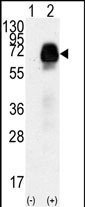
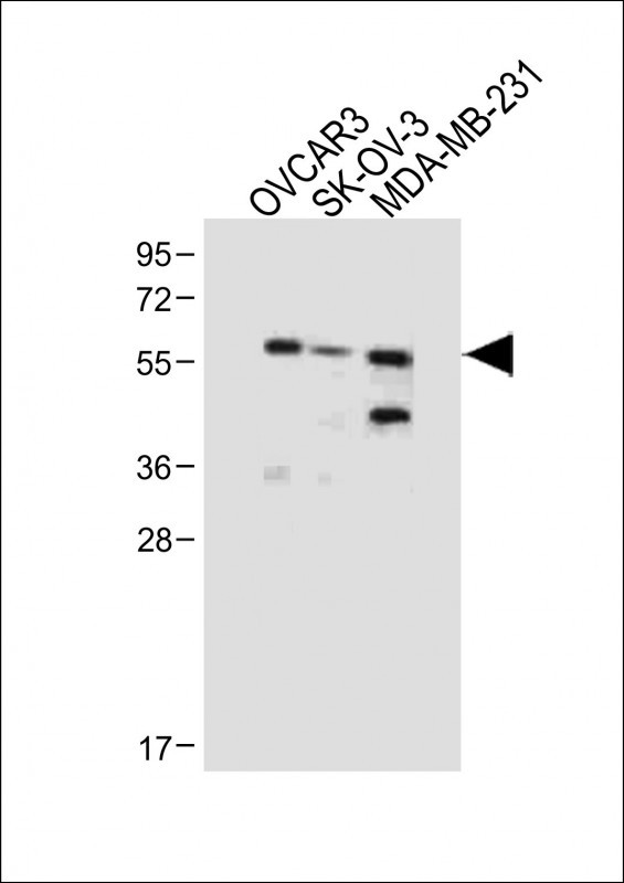
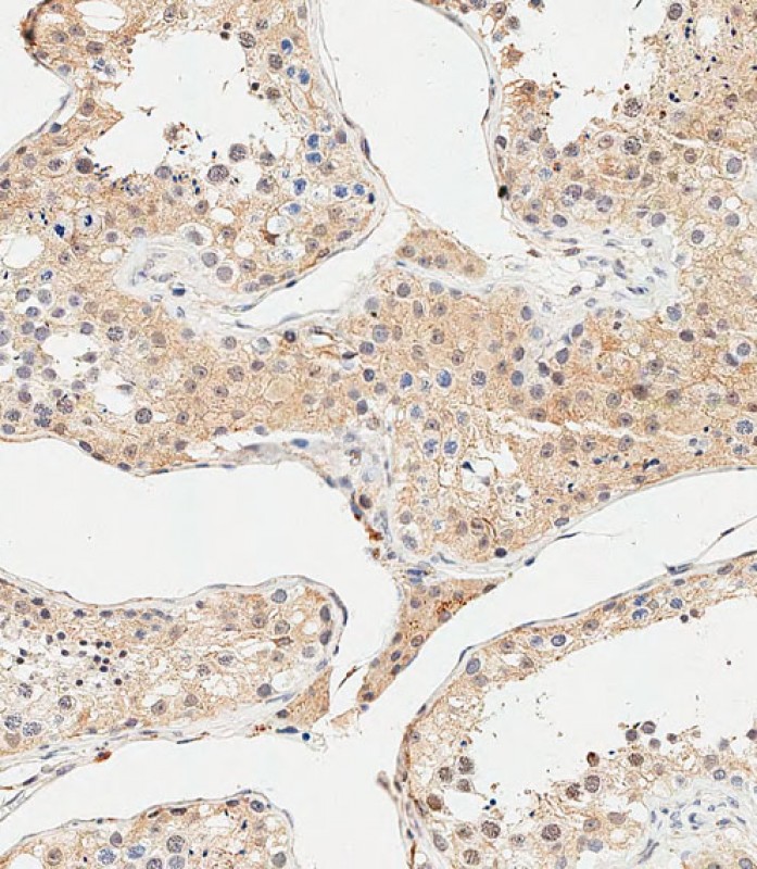
AMHR2 Antibody (N-term R80)
Purified Rabbit Polyclonal Antibody (Pab)
- SPECIFICATION
- CITATIONS
- PROTOCOLS
- BACKGROUND

Application
| IHC-P-Leica, WB, E |
|---|---|
| Primary Accession | Q16671 |
| Other Accession | Q8K592 |
| Reactivity | Human |
| Predicted | Mouse |
| Host | Rabbit |
| Clonality | Polyclonal |
| Isotype | Rabbit IgG |
| Calculated MW | 62750 Da |
| Antigen Region | 65-91 aa |
| Gene ID | 269 |
|---|---|
| Other Names | Anti-Muellerian hormone type-2 receptor, Anti-Muellerian hormone type II receptor, AMH type II receptor, MIS type II receptor, MISRII, MRII, AMHR2, AMHR, MISR2 |
| Target/Specificity | This AMHR2 antibody is generated from rabbits immunized with a KLH conjugated synthetic peptide between 65-91 amino acids from the N-terminal region of human AMHR2. |
| Dilution | IHC-P-Leica~~1:500 WB~~1:1000 E~~Use at an assay dependent concentration. |
| Format | Purified polyclonal antibody supplied in PBS with 0.09% (W/V) sodium azide. This antibody is purified through a protein A column, followed by peptide affinity purification. |
| Storage | Maintain refrigerated at 2-8°C for up to 2 weeks. For long term storage store at -20°C in small aliquots to prevent freeze-thaw cycles. |
| Precautions | AMHR2 Antibody (N-term R80) is for research use only and not for use in diagnostic or therapeutic procedures. |
| Name | AMHR2 |
|---|---|
| Synonyms | AMHR, MISR2 |
| Function | On ligand binding, forms a receptor complex consisting of two type II and two type I transmembrane serine/threonine kinases. Type II receptors phosphorylate and activate type I receptors which autophosphorylate, then bind and activate SMAD transcriptional regulators. Receptor for anti-Muellerian hormone. |
| Cellular Location | Membrane; Single-pass type I membrane protein. |

Thousands of laboratories across the world have published research that depended on the performance of antibodies from Abcepta to advance their research. Check out links to articles that cite our products in major peer-reviewed journals, organized by research category.
info@abcepta.com, and receive a free "I Love Antibodies" mug.

Provided below are standard protocols that you may find useful for product applications.
Background
The AMH receptor (AMHR or AMHR2) is a serine/threonine kinase with a single transmembrane domain belonging to the family of type II receptors for TGF-beta-related proteins. Anti-Mullerian hormone (AMH) and its receptor are involved in the regression of Mullerian ducts in male fetuses. Male sex differentiation is mediated by 2 discrete hormones produced by the fetal testis. Testosterone, produced by Leydig cells, virilizes the external genitalia and promotes prostatic growth; anti-Mullerian hormone (AMH) results in regression of Mullerian ducts which would otherwise differentiate into the uterus and fallopian tubes.
References
Picard, J.Y., et al., J. Soc. Biol. 196(3):217-221 (2002).
Teixeira, J., et al., Endocr. Rev. 22(5):657-674 (2001).
Imbeaud, S., et al., Nat. Genet. 11(4):382-388 (1995).
Visser, J.A., et al., Biochem. Biophys. Res. Commun. 215(3):1029-1036 (1995).
Sinisi, A.A., et al., J. Endocrinol. Invest. 26 (3 Suppl), 23-28 (2003).
If you have used an Abcepta product and would like to share how it has performed, please click on the "Submit Review" button and provide the requested information. Our staff will examine and post your review and contact you if needed.
If you have any additional inquiries please email technical services at tech@abcepta.com.
Ordering Information
Other Products
Shipping Information
