


 Foundational characteristics of cancer include proliferation, angiogenesis, migration, evasion of apoptosis, and cellular immortality. Find key markers for these cellular processes and antibodies to detect them.
 The SUMOplot™ Analysis Program predicts and scores sumoylation sites in your protein. SUMOylation is a post-translational modification involved in various cellular processes, such as nuclear-cytosolic transport, transcriptional regulation, apoptosis, protein stability, response to stress, and progression through the cell cycle.
 The Autophagy Receptor Motif Plotter predicts and scores autophagy receptor binding sites in your protein. Identifying proteins connected to this pathway is critical to understanding the role of autophagy in physiological as well as pathological processes such as development, differentiation, neurodegenerative diseases, stress, infection, and cancer.
NME3 Antibody (Center)
Purified Rabbit Polyclonal Antibody (Pab)
- SPECIFICATION
- CITATIONS
- PROTOCOLS
- BACKGROUND

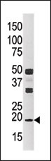
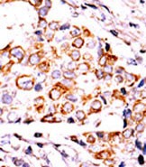
Application
| WB, IHC-P, E |
|---|---|
| Primary Accession | Q13232 |
| Reactivity | Human |
| Host | Rabbit |
| Clonality | Polyclonal |
| Isotype | Rabbit IgG |
| Calculated MW | 19015 Da |
| Antigen Region | 51-81 aa |
| Gene ID | 4832 |
|---|---|
| Other Names | Nucleoside diphosphate kinase 3, NDK 3, NDP kinase 3, DR-nm23, Nucleoside diphosphate kinase C, NDPKC, nm23-H3, NME3 |
| Target/Specificity | This NME3 antibody is generated from rabbits immunized with a KLH conjugated synthetic peptide between 51-81 amino acids from the Central region of human NME3. |
| Dilution | WB~~1:1000 IHC-P~~1:50~100 E~~Use at an assay dependent concentration. |
| Format | Purified polyclonal antibody supplied in PBS with 0.09% (W/V) sodium azide. This antibody is prepared by Saturated Ammonium Sulfate (SAS) precipitation followed by dialysis against PBS. |
| Storage | Maintain refrigerated at 2-8°C for up to 2 weeks. For long term storage store at -20°C in small aliquots to prevent freeze-thaw cycles. |
| Precautions | NME3 Antibody (Center) is for research use only and not for use in diagnostic or therapeutic procedures. |
| Name | NME3 (HGNC:7851) |
|---|---|
| Function | Catalyzes the phosphorylation of ribonucleosides and deoxyribonucleoside diphosphates, other than ATP, into the corresponding triphosphates with ATP as the major phosphate donor (PubMed:11277919, PubMed:30587587). The ATP gamma phosphate is transferred to the nucleoside diphosphate beta phosphate via a ping- pong mechanism, using a phosphorylated active-site intermediate. Through the catalyzed exchange of gamma-phosphate between di- and triphosphonucleosides participates in regulation of intracellular nucleotide homeostasis (PubMed:11277919, PubMed:30587587). Inhibits granulocyte differentiation (PubMed:7638209). May be required for ciliary function during renal development (By similarity). |
| Cellular Location | Mitochondrion outer membrane; Peripheral membrane protein. Cytoplasm Cytoplasm, cytoskeleton, cilium basal body {ECO:0000250|UniProtKB:Q9WV85} |

Thousands of laboratories across the world have published research that depended on the performance of antibodies from Abcepta to advance their research. Check out links to articles that cite our products in major peer-reviewed journals, organized by research category.
info@abcepta.com, and receive a free "I Love Antibodies" mug.

Provided below are standard protocols that you may find useful for product applications.
Background
NME3 mRNA is preferentially expressed at early stages of myeloid differentiation of highly purified CD34(+) cells. Its constitutive expression in a myeloid precursor line, which is growth-factor dependent for both proliferation and differentiation, results in inhibition of granulocytic differentiation induced by granulocyte colony-stimulating factor and causes apoptotic cell death. These results appear consistent with a role for the NME3 gene in normal hematopoiesis and raise the possibility that its overexpression contributes to differentiation arrest, a feature of blastic transformation in chronic myelogenous leukemia.
References
Negroni, A., et al., Cell Death Differ. 7(9):843-850 (2000).
Martinez, R., et al., Cancer Res. 57(6):1180-1187 (1997).
Venturelli, D., et al., Proc. Natl. Acad. Sci. U.S.A. 92(16):7435-7439 (1995).
If you have used an Abcepta product and would like to share how it has performed, please click on the "Submit Review" button and provide the requested information. Our staff will examine and post your review and contact you if needed.
If you have any additional inquiries please email technical services at tech@abcepta.com.
Ordering Information
Other Products
Shipping Information
