


 Foundational characteristics of cancer include proliferation, angiogenesis, migration, evasion of apoptosis, and cellular immortality. Find key markers for these cellular processes and antibodies to detect them.
 The SUMOplot™ Analysis Program predicts and scores sumoylation sites in your protein. SUMOylation is a post-translational modification involved in various cellular processes, such as nuclear-cytosolic transport, transcriptional regulation, apoptosis, protein stability, response to stress, and progression through the cell cycle.
 The Autophagy Receptor Motif Plotter predicts and scores autophagy receptor binding sites in your protein. Identifying proteins connected to this pathway is critical to understanding the role of autophagy in physiological as well as pathological processes such as development, differentiation, neurodegenerative diseases, stress, infection, and cancer.
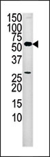
PANK2 Antibody (Center)
Purified Rabbit Polyclonal Antibody (Pab)
- SPECIFICATION
- CITATIONS
- PROTOCOLS
- BACKGROUND

Application
| WB, E |
|---|---|
| Primary Accession | Q9BZ23 |
| Reactivity | Human |
| Host | Rabbit |
| Clonality | Polyclonal |
| Isotype | Rabbit IgG |
| Calculated MW | 62681 Da |
| Antigen Region | 180-210 aa |
| Gene ID | 80025 |
|---|---|
| Other Names | Pantothenate kinase 2, mitochondrial, hPanK2, Pantothenic acid kinase 2, PANK2, C20orf48 |
| Target/Specificity | This PANK2 antibody is generated from rabbits immunized with a KLH conjugated synthetic peptide between 180-210 amino acids from the Central region of human PANK2. |
| Dilution | WB~~1:1000 E~~Use at an assay dependent concentration. |
| Format | Purified polyclonal antibody supplied in PBS with 0.09% (W/V) sodium azide. This antibody is prepared by Saturated Ammonium Sulfate (SAS) precipitation followed by dialysis against PBS. |
| Storage | Maintain refrigerated at 2-8°C for up to 2 weeks. For long term storage store at -20°C in small aliquots to prevent freeze-thaw cycles. |
| Precautions | PANK2 Antibody (Center) is for research use only and not for use in diagnostic or therapeutic procedures. |
| Name | PANK2 |
|---|---|
| Synonyms | C20orf48 |
| Function | [Isoform 1]: Mitochondrial isoform that catalyzes the phosphorylation of pantothenate to generate 4'-phosphopantothenate in the first and rate-determining step of coenzyme A (CoA) synthesis (PubMed:15659606, PubMed:16272150, PubMed:17242360, PubMed:17825826). Required for angiogenic activity of umbilical vein of endothelial cells (HUVEC) (PubMed:30221726). |
| Cellular Location | [Isoform 1]: Mitochondrion. Mitochondrion intermembrane space. Nucleus Note=Localizes predominantly to the mitochondria and to a lesser extent to the nucleus. Found in both the mitochondria and the nucleus throughout the cell cycle, with the exception of the G2/M phase when it is restricted to mitochdondria. [Isoform 3]: Cytoplasm {ECO:0000269|PubMed:12554685, ECO:0000305} |
| Tissue Location | Expressed in the brain (at protein level) (PubMed:15659606, PubMed:17825826). Ubiquitous (PubMed:11479594) Highly expressed in the testis (PubMed:17825826). Expressed in the umbilical vein endothelial cells (HUVEC) (PubMed:30221726) |

Thousands of laboratories across the world have published research that depended on the performance of antibodies from Abcepta to advance their research. Check out links to articles that cite our products in major peer-reviewed journals, organized by research category.
info@abcepta.com, and receive a free "I Love Antibodies" mug.

Provided below are standard protocols that you may find useful for product applications.
Background
Pantothenate kinase is an essential regulatory enzyme in CoA biosynthesis, catalyzing the cytosolic phosphorylation of pantothenate (vitamin B5), N-pantothenoylcysteine, and pantetheine. CoA is the major acyl carrier, playing a central role in intermediary and fatty acid metabolism. In both yeast and fly, each with only 1 pantothenate kinase gene, the null mutant is inviable. Mutations in PANK2 are the cause of pantothenate kinase-associated neurodegeneration (PKAN), formerly known as Hallervorden-Spatz syndrome (HSS). PKAN is an autosomal recessive neurodegenerative disorder associated with iron accumulation in the brain. Mutations in PANK2 are the cause of hypoprebetalipoproteinemia, acanthocytosis, retinitis pigmentosa, and pallidal degeneration (HARP).
References
Neurology 58: 1673-1674, 2002.
Hum. Molec. Genet. 12: 321-327, 2003.
Neurology 61: 1423-1426, 2003.
Neurology 64: 1810-1812, 2005.
Nature Genet. 28: 345-349, 2001.
If you have used an Abcepta product and would like to share how it has performed, please click on the "Submit Review" button and provide the requested information. Our staff will examine and post your review and contact you if needed.
If you have any additional inquiries please email technical services at tech@abcepta.com.
Ordering Information
Other Products
Shipping Information
