


 Foundational characteristics of cancer include proliferation, angiogenesis, migration, evasion of apoptosis, and cellular immortality. Find key markers for these cellular processes and antibodies to detect them.
 The SUMOplot™ Analysis Program predicts and scores sumoylation sites in your protein. SUMOylation is a post-translational modification involved in various cellular processes, such as nuclear-cytosolic transport, transcriptional regulation, apoptosis, protein stability, response to stress, and progression through the cell cycle.
 The Autophagy Receptor Motif Plotter predicts and scores autophagy receptor binding sites in your protein. Identifying proteins connected to this pathway is critical to understanding the role of autophagy in physiological as well as pathological processes such as development, differentiation, neurodegenerative diseases, stress, infection, and cancer.
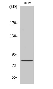
Pinin Polyclonal Antibody
- SPECIFICATION
- CITATIONS
- PROTOCOLS
- BACKGROUND

Application
| WB |
|---|---|
| Primary Accession | Q9H307 |
| Reactivity | Human, Mouse |
| Host | Rabbit |
| Clonality | Polyclonal |
| Calculated MW | 81628 Da |
| Gene ID | 5411 |
|---|---|
| Other Names | PNN; DRS; MEMA; Pinin; 140 kDa nuclear and cell adhesion-related phosphoprotein; Desmosome-associated protein; Domain-rich serine protein; DRS protein; DRSP; Melanoma metastasis clone A protein; Nuclear protein SDK3; SR-like protein |
| Dilution | WB~~Western Blot: 1/500 - 1/2000. ELISA: 1/20000. Not yet tested in other applications. |
| Format | Liquid in PBS containing 50% glycerol, 0.5% BSA and 0.09% (W/V) sodium azide. |
| Storage Conditions | -20℃ |
| Name | PNN |
|---|---|
| Synonyms | DRS, MEMA |
| Function | Transcriptional activator binding to the E-box 1 core sequence of the E-cadherin promoter gene; the core-binding sequence is 5'CAGGTG-3'. Capable of reversing CTBP1-mediated transcription repression. Auxiliary component of the splicing-dependent multiprotein exon junction complex (EJC) deposited at splice junction on mRNAs. The EJC is a dynamic structure consisting of core proteins and several peripheral nuclear and cytoplasmic associated factors that join the complex only transiently either during EJC assembly or during subsequent mRNA metabolism. Participates in the regulation of alternative pre-mRNA splicing. Associates to spliced mRNA within 60 nt upstream of the 5'-splice sites. Component of the PSAP complex which binds RNA in a sequence-independent manner and is proposed to be recruited to the EJC prior to or during the splicing process and to regulate specific excision of introns in specific transcription subsets. Involved in the establishment and maintenance of epithelia cell-cell adhesion. Potential tumor suppressor for renal cell carcinoma. |
| Cellular Location | Nucleus speckle. Cell junction, desmosome. Note=Cell-cell contact area, predominantly desmosome of intercellular adherens junction. Not a nucleocytoplasmic shuttling protein |
| Tissue Location | Expressed in placenta, lung, liver, kidney, pancreas, spleen, thymus, prostate, testis, ovary, small intestine, colon, heart, epidermis, esophagus, brain and smooth and skeletal muscle. Expressed strongly in melanoma metastasis lesions and advanced primary tumors. |

Thousands of laboratories across the world have published research that depended on the performance of antibodies from Abcepta to advance their research. Check out links to articles that cite our products in major peer-reviewed journals, organized by research category.
info@abcepta.com, and receive a free "I Love Antibodies" mug.

Provided below are standard protocols that you may find useful for product applications.
Background
Transcriptional activator binding to the E-box 1 core sequence of the E-cadherin promoter gene; the core-binding sequence is 5'CAGGTG-3'. Capable of reversing CTBP1-mediated transcription repression. Auxiliary component of the splicing- dependent multiprotein exon junction complex (EJC) deposited at splice junction on mRNAs. The EJC is a dynamic structure consisting of core proteins and several peripheral nuclear and cytoplasmic associated factors that join the complex only transiently either during EJC assembly or during subsequent mRNA metabolism. Participates in the regulation of alternative pre-mRNA splicing. Associates to spliced mRNA within 60 nt upstream of the 5'-splice sites. Component of the PSAP complex which binds RNA in a sequence-independent manner and is proposed to be recruited to the EJC prior to or during the splicing process and to regulate specific excision of introns in specific transcription subsets. Involved in the establishment and maintenance of epithelia cell- cell adhesion. Potential tumor suppressor for renal cell carcinoma.
If you have used an Abcepta product and would like to share how it has performed, please click on the "Submit Review" button and provide the requested information. Our staff will examine and post your review and contact you if needed.
If you have any additional inquiries please email technical services at tech@abcepta.com.
Ordering Information
Other Products
Shipping Information
