


 Foundational characteristics of cancer include proliferation, angiogenesis, migration, evasion of apoptosis, and cellular immortality. Find key markers for these cellular processes and antibodies to detect them.
 The SUMOplot™ Analysis Program predicts and scores sumoylation sites in your protein. SUMOylation is a post-translational modification involved in various cellular processes, such as nuclear-cytosolic transport, transcriptional regulation, apoptosis, protein stability, response to stress, and progression through the cell cycle.
 The Autophagy Receptor Motif Plotter predicts and scores autophagy receptor binding sites in your protein. Identifying proteins connected to this pathway is critical to understanding the role of autophagy in physiological as well as pathological processes such as development, differentiation, neurodegenerative diseases, stress, infection, and cancer.
Renin Receptor Polyclonal Antibody
- SPECIFICATION
- CITATIONS
- PROTOCOLS
- BACKGROUND

Application
| WB, IHC-P, IF |
|---|---|
| Primary Accession | O75787 |
| Reactivity | Human, Mouse, Rat |
| Host | Rabbit |
| Clonality | Polyclonal |
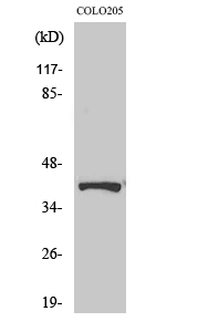
| Calculated MW | 39008 Da |
| Gene ID | 10159 |
|---|---|
| Other Names | ATP6AP2; ATP6IP2; CAPER; ELDF10; HT028; MSTP009; PSEC0072; Renin receptor; ATPase H(+)-transporting lysosomal accessory protein 2; ATPase H(+)-transporting lysosomal-interacting protein 2; ER-localized type I transmembrane adaptor; Embryoni |
| Dilution | WB~~Western Blot: 1/500 - 1/2000. Immunohistochemistry: 1/100 - 1/300. Immunofluorescence: 1/200 - 1/1000. ELISA: 1/20000. Not yet tested in other applications. IHC-P~~N/A IF~~1:50~200 |
| Format | Liquid in PBS containing 50% glycerol, 0.5% BSA and 0.09% (W/V) sodium azide. |
| Storage Conditions | -20℃ |
| Name | ATP6AP2 (HGNC:18305) |
|---|---|
| Function | Multifunctional protein which functions as a renin, prorenin cellular receptor and is involved in the assembly of the lysosomal proton-transporting V-type ATPase (V-ATPase) and the acidification of the endo-lysosomal system (PubMed:12045255, PubMed:29127204, PubMed:30374053, PubMed:32276428). May mediate renin-dependent cellular responses by activating ERK1 and ERK2 (PubMed:12045255). By increasing the catalytic efficiency of renin in AGT/angiotensinogen conversion to angiotensin I, may also play a role in the renin-angiotensin system (RAS) (PubMed:12045255). Through its function in V-type ATPase (v- ATPase) assembly and acidification of the lysosome it regulates protein degradation and may control different signaling pathways important for proper brain development, synapse morphology and synaptic transmission (By similarity). |
| Cellular Location | Endoplasmic reticulum membrane; Single-pass type I membrane protein. Lysosome membrane; Single- pass type I membrane protein. Cytoplasmic vesicle, autophagosome membrane {ECO:0000250|UniProtKB:Q9CYN9}; Single-pass type I membrane protein. Cell projection, dendritic spine membrane {ECO:0000250|UniProtKB:Q9CYN9}; Single-pass type I membrane protein. Cell projection, axon {ECO:0000250|UniProtKB:Q9CYN9}. Endosome membrane {ECO:0000250|UniProtKB:Q9CYN9}; Single-pass type I membrane protein. Cytoplasmic vesicle, clathrin-coated vesicle membrane {ECO:0000250|UniProtKB:Q6AXS4}; Single-pass type I membrane protein. Cytoplasmic vesicle, secretory vesicle, synaptic vesicle membrane {ECO:0000250|UniProtKB:Q6AXS4}; Single-pass type I membrane protein |
| Tissue Location | Expressed in brain, heart, placenta, liver, kidney and pancreas. Barely detectable in lung and skeletal muscles. In the kidney cortex it is restricted to the mesangium of glomeruli. In the coronary and kidney artery it is expressed in the subendothelium, associated to smooth muscles where it colocalizes with REN. Expressed in vascular structures and by syncytiotrophoblast cells in the mature fetal placenta. |

Thousands of laboratories across the world have published research that depended on the performance of antibodies from Abcepta to advance their research. Check out links to articles that cite our products in major peer-reviewed journals, organized by research category.
info@abcepta.com, and receive a free "I Love Antibodies" mug.

Provided below are standard protocols that you may find useful for product applications.
Background
Functions as a renin and prorenin cellular receptor. May mediate renin-dependent cellular responses by activating ERK1 and ERK2. By increasing the catalytic efficiency of renin in AGT/angiotensinogen conversion to angiotensin I, it may also play a role in the renin-angiotensin system (RAS).
If you have used an Abcepta product and would like to share how it has performed, please click on the "Submit Review" button and provide the requested information. Our staff will examine and post your review and contact you if needed.
If you have any additional inquiries please email technical services at tech@abcepta.com.
Ordering Information
Other Products
Shipping Information
