


 Foundational characteristics of cancer include proliferation, angiogenesis, migration, evasion of apoptosis, and cellular immortality. Find key markers for these cellular processes and antibodies to detect them.
 The SUMOplot™ Analysis Program predicts and scores sumoylation sites in your protein. SUMOylation is a post-translational modification involved in various cellular processes, such as nuclear-cytosolic transport, transcriptional regulation, apoptosis, protein stability, response to stress, and progression through the cell cycle.
 The Autophagy Receptor Motif Plotter predicts and scores autophagy receptor binding sites in your protein. Identifying proteins connected to this pathway is critical to understanding the role of autophagy in physiological as well as pathological processes such as development, differentiation, neurodegenerative diseases, stress, infection, and cancer.
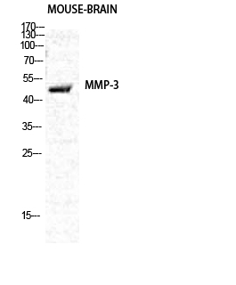
MMP-3 Polyclonal Antibody
.jpg)
- SPECIFICATION
- CITATIONS
- PROTOCOLS
- BACKGROUND

Application
| WB, IHC-P, IF |
|---|---|
| Primary Accession | P08254 |
| Reactivity | Human, Mouse, Rat |
| Host | Rabbit |
| Clonality | Polyclonal |
| Calculated MW | 53977 Da |
| Gene ID | 4314 |
|---|---|
| Other Names | MMP3; STMY1; Stromelysin-1; SL-1; Matrix metalloproteinase-3; MMP-3; Transin-1 |
| Dilution | WB~~Western Blot: 1/500 - 1/2000. Immunohistochemistry: 1/100 - 1/300. Immunofluorescence: 1/200 - 1/1000. ELISA: 1/10000. Not yet tested in other applications. IHC-P~~N/A IF~~1:50~200 |
| Format | Liquid in PBS containing 50% glycerol, 0.5% BSA and 0.09% (W/V) sodium azide. |
| Storage Conditions | -20℃ |
| Name | MMP3 |
|---|---|
| Synonyms | STMY1 |
| Function | Metalloproteinase with a rather broad substrate specificity that can degrade fibronectin, laminin, gelatins of type I, III, IV, and V; collagens III, IV, X, and IX, and cartilage proteoglycans. Activates different molecules including growth factors, plasminogen or other matrix metalloproteinases such as MMP9 (PubMed:11029580, PubMed:1371271). Once released into the extracellular matrix (ECM), the inactive pro-enzyme is activated by the plasmin cascade signaling pathway (PubMed:2383557). Also acts intracellularly (PubMed:22265821). For example, in dopaminergic neurons, gets activated by the serine protease HTRA2 upon stress and plays a pivotal role in DA neuronal degeneration by mediating microglial activation and alpha- synuclein/SNCA cleavage (PubMed:21330369). In addition, plays a role in immune response and possesses antiviral activity against various viruses such as vesicular stomatitis virus, influenza A virus (H1N1) and human herpes virus 1 (PubMed:35940311). Mechanistically, translocates from the cytoplasm into the cell nucleus upon virus infection to influence NF-kappa-B activities (PubMed:35940311). |
| Cellular Location | Secreted, extracellular space, extracellular matrix. Nucleus. Cytoplasm |

Thousands of laboratories across the world have published research that depended on the performance of antibodies from Abcepta to advance their research. Check out links to articles that cite our products in major peer-reviewed journals, organized by research category.
info@abcepta.com, and receive a free "I Love Antibodies" mug.

Provided below are standard protocols that you may find useful for product applications.
Background
Can degrade fibronectin, laminin, gelatins of type I, III, IV, and V; collagens III, IV, X, and IX, and cartilage proteoglycans. Activates procollagenase.
If you have used an Abcepta product and would like to share how it has performed, please click on the "Submit Review" button and provide the requested information. Our staff will examine and post your review and contact you if needed.
If you have any additional inquiries please email technical services at tech@abcepta.com.
Ordering Information
Other Products
Shipping Information
