


 Foundational characteristics of cancer include proliferation, angiogenesis, migration, evasion of apoptosis, and cellular immortality. Find key markers for these cellular processes and antibodies to detect them.
 The SUMOplot™ Analysis Program predicts and scores sumoylation sites in your protein. SUMOylation is a post-translational modification involved in various cellular processes, such as nuclear-cytosolic transport, transcriptional regulation, apoptosis, protein stability, response to stress, and progression through the cell cycle.
 The Autophagy Receptor Motif Plotter predicts and scores autophagy receptor binding sites in your protein. Identifying proteins connected to this pathway is critical to understanding the role of autophagy in physiological as well as pathological processes such as development, differentiation, neurodegenerative diseases, stress, infection, and cancer.
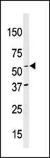
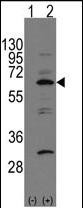
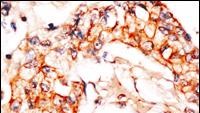
NAE1 (APPBP1) Antibody (C-term)
Purified Rabbit Polyclonal Antibody (Pab)
- SPECIFICATION
- CITATIONS
- PROTOCOLS
- BACKGROUND

Application
| IHC-P, WB, E |
|---|---|
| Primary Accession | Q13564 |
| Other Accession | Q9Z1A5, Q8VBW6, Q4R3L6, NP_003896 |
| Reactivity | Human |
| Predicted | Monkey, Mouse, Rat |
| Host | Rabbit |
| Clonality | Polyclonal |
| Isotype | Rabbit IgG |
| Calculated MW | 60246 Da |
| Antigen Region | 430-459 aa |
| Gene ID | 8883 |
|---|---|
| Other Names | NEDD8-activating enzyme E1 regulatory subunit, Amyloid beta precursor protein-binding protein 1, 59 kDa, APP-BP1, Amyloid protein-binding protein 1, Proto-oncogene protein 1, NAE1, APPBP1 |
| Target/Specificity | This NAE1 (APPBP1) antibody is generated from rabbits immunized with a KLH conjugated synthetic peptide between 430-459 amino acids from the C-terminal region of human NAE1 (APPBP1). |
| Dilution | IHC-P~~1:50~100 WB~~1:1000 E~~Use at an assay dependent concentration. |
| Format | Purified polyclonal antibody supplied in PBS with 0.09% (W/V) sodium azide. This antibody is purified through a protein G column, followed by dialysis against PBS. |
| Storage | Maintain refrigerated at 2-8°C for up to 2 weeks. For long term storage store at -20°C in small aliquots to prevent freeze-thaw cycles. |
| Precautions | NAE1 (APPBP1) Antibody (C-term) is for research use only and not for use in diagnostic or therapeutic procedures. |
| Name | NAE1 |
|---|---|
| Synonyms | APPBP1 |
| Function | Regulatory subunit of the dimeric UBA3-NAE1 E1 enzyme. E1 activates NEDD8 by first adenylating its C-terminal glycine residue with ATP, thereafter linking this residue to the side chain of the catalytic cysteine, yielding a NEDD8-UBA3 thioester and free AMP. E1 finally transfers NEDD8 to the catalytic cysteine of UBE2M. Necessary for cell cycle progression through the S-M checkpoint. Overexpression of NAE1 causes apoptosis through deregulation of NEDD8 conjugation. The covalent attachment of NEDD8 to target proteins is known as 'neddylation' and the process is involved in the regulation of cell growth, viability and development. |
| Cellular Location | Cell membrane. Note=Colocalizes with APP in lipid rafts |
| Tissue Location | Ubiquitous in fetal tissues. Expressed throughout the adult brain. |

Thousands of laboratories across the world have published research that depended on the performance of antibodies from Abcepta to advance their research. Check out links to articles that cite our products in major peer-reviewed journals, organized by research category.
info@abcepta.com, and receive a free "I Love Antibodies" mug.

Provided below are standard protocols that you may find useful for product applications.
Background
APPBP1 binds to the beta-amyloid precursor protein, a cell surface protein with signal-transducing properties thought to play a role in the pathogenesis of Alzheimer's disease. In addition, this protein can form a heterodimer with UBE1C and bind and activate NEDD8, a ubiquitin-like protein. APPB1 is required for cell cycle progression through the S/M checkpoint.
References
Chen,Y., J. Cell Biol. 163 (1), 27-33 (2003)
Chen,Y., J. Neurochem. 85 (3), 801-809 (2003)
Walden,H., Nature 422 (6929), 330-334 (2003)
Chow,N., J. Biol. Chem. 271 (19), 11339-11346 (1996)
If you have used an Abcepta product and would like to share how it has performed, please click on the "Submit Review" button and provide the requested information. Our staff will examine and post your review and contact you if needed.
If you have any additional inquiries please email technical services at tech@abcepta.com.
Ordering Information
Other Products
Shipping Information
