


 Foundational characteristics of cancer include proliferation, angiogenesis, migration, evasion of apoptosis, and cellular immortality. Find key markers for these cellular processes and antibodies to detect them.
 The SUMOplot™ Analysis Program predicts and scores sumoylation sites in your protein. SUMOylation is a post-translational modification involved in various cellular processes, such as nuclear-cytosolic transport, transcriptional regulation, apoptosis, protein stability, response to stress, and progression through the cell cycle.
 The Autophagy Receptor Motif Plotter predicts and scores autophagy receptor binding sites in your protein. Identifying proteins connected to this pathway is critical to understanding the role of autophagy in physiological as well as pathological processes such as development, differentiation, neurodegenerative diseases, stress, infection, and cancer.
CD85g Polyclonal Antibody
.jpg)

- SPECIFICATION
- CITATIONS
- PROTOCOLS
- BACKGROUND

Application
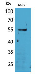
| WB, IHC-P |
|---|---|
| Primary Accession | P59901 |
| Reactivity | Human |
| Host | Rabbit |
| Clonality | Polyclonal |
| Calculated MW | 55181 Da |
| Gene ID | 23547 |
|---|---|
| Other Names | LILRA4; ILT7; Leukocyte immunoglobulin-like receptor subfamily A member 4; CD85 antigen-like family member G; Immunoglobulin-like transcript 7; ILT-7; CD85g |
| Dilution | WB~~Western Blot: 1/500 - 1/2000. IHC-p: 1:100-300 ELISA: 1/20000. Not yet tested in other applications. IHC-P~~N/A |
| Format | Liquid in PBS containing 50% glycerol, 0.5% BSA and 0.09% (W/V) sodium azide. |
| Storage Conditions | -20℃ |
| Name | LILRA4 (HGNC:15503) |
|---|---|
| Function | Functions coreceptor to limit the innate immune responses to viral infections; signaling occurs via FCER1G (PubMed:16735691, PubMed:19564354). Down-regulates the production of IFNA1, IFNA2, IFNA4, IFNB1 and TNF by plasmacytoid dendritic cells that have been exposed to influenza virus or cytidine-phosphate-guanosine (CpG) dinucleotides, indicating it functions as a negative regulator of TLR7 and TLR9 signaling cascades (PubMed:16735691, PubMed:19564354, PubMed:24586760). Down-regulates interferon production in response to interaction with BST2 on HIV-1 infected cells (PubMed:26172439). Activates a signaling cascade in complex with FCER1G that results in phosphorylation of Src family and Syk kinases and thereby triggers mobilization of intracellular Ca(2+) (PubMed:16735691, PubMed:19564354). Does not interfere with the differentiation of plasmacytoid dendritic cells into antigen-presenting cells (PubMed:24586760). |
| Cellular Location | Cell membrane; Single-pass type I membrane protein |
| Tissue Location | Detected on plasmacytoid dendritic cells (at protein level). Detected on plasmacytoid dendritic cells, but not on monocytes or B cells. |

Thousands of laboratories across the world have published research that depended on the performance of antibodies from Abcepta to advance their research. Check out links to articles that cite our products in major peer-reviewed journals, organized by research category.
info@abcepta.com, and receive a free "I Love Antibodies" mug.

Provided below are standard protocols that you may find useful for product applications.
Background
Functions coreceptor to limit the innate immune responses to viral infections; signaling occurs via FCER1G (PubMed:16735691, PubMed:19564354). Down-regulates the production of IFNA1, IFNA2, IFNA4, IFNB1 and TNF by plasmacytoid dendritic cells that have been exposed to influenza virus or cytidine- phosphate-guanosine (CpG) dinucleotides, indicating it functions as negative regulator of TLR7 and TLR9 signaling cascades (PubMed:16735691, PubMed:19564354, PubMed:24586760). Down- regulates interferon production in response to interaction with BST2 on HIV-1 infected cells (PubMed:26172439). Activates a signaling cascade in complex with FCER1G that results in phosphorylation of Src family and Syk kinases and thereby triggers mobilization of intracellular Ca(2+) (PubMed:16735691, PubMed:19564354). Does not interfere with the differentiation of plasmacytoid dendritic cells into antigen-presenting cells (PubMed:24586760).
If you have used an Abcepta product and would like to share how it has performed, please click on the "Submit Review" button and provide the requested information. Our staff will examine and post your review and contact you if needed.
If you have any additional inquiries please email technical services at tech@abcepta.com.
Ordering Information
Other Products
Shipping Information
