


 Foundational characteristics of cancer include proliferation, angiogenesis, migration, evasion of apoptosis, and cellular immortality. Find key markers for these cellular processes and antibodies to detect them.
 The SUMOplot™ Analysis Program predicts and scores sumoylation sites in your protein. SUMOylation is a post-translational modification involved in various cellular processes, such as nuclear-cytosolic transport, transcriptional regulation, apoptosis, protein stability, response to stress, and progression through the cell cycle.
 The Autophagy Receptor Motif Plotter predicts and scores autophagy receptor binding sites in your protein. Identifying proteins connected to this pathway is critical to understanding the role of autophagy in physiological as well as pathological processes such as development, differentiation, neurodegenerative diseases, stress, infection, and cancer.
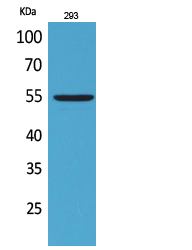
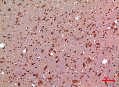
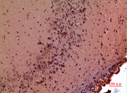
GDF-5 Polyclonal Antibody
.jpg)
- SPECIFICATION
- CITATIONS
- PROTOCOLS
- BACKGROUND

Application
| WB, IHC-P |
|---|---|
| Primary Accession | P43026 |
| Reactivity | Human, Mouse, Rat |
| Host | Rabbit |
| Clonality | Polyclonal |
| Calculated MW | 55395 Da |
| Gene ID | 8200 |
|---|---|
| Other Names | GDF5; CDMP1; Growth/differentiation factor 5; GDF-5; Cartilage-derived morphogenetic protein 1; CDMP-1; Radotermin |
| Dilution | WB~~Western Blot: 1/500 - 1/2000. IHC-p: 1/100-1/300. ELISA: 1/20000. Not yet tested in other applications. IHC-P~~N/A |
| Format | Liquid in PBS containing 50% glycerol, 0.5% BSA and 0.09% (W/V) sodium azide. |
| Storage Conditions | -20℃ |
| Name | GDF5 |
|---|---|
| Synonyms | BMP14, CDMP1 |
| Function | Growth factor involved in bone and cartilage formation. During cartilage development regulates differentiation of chondrogenic tissue through two pathways. Firstly, positively regulates differentiation of chondrogenic tissue through its binding of high affinity with BMPR1B and of less affinity with BMPR1A, leading to induction of SMAD1-SMAD5-SMAD8 complex phosphorylation and then SMAD protein signaling transduction (PubMed:15530414, PubMed:21976273, PubMed:24098149, PubMed:25092592). Secondly, negatively regulates chondrogenic differentiation through its interaction with NOG (PubMed:21976273). Required to prevent excessive muscle loss upon denervation. This function requires SMAD4 and is mediated by phosphorylated SMAD1/5/8 (By similarity). Binds bacterial lipopolysaccharide (LPS) and mediates LPS-induced inflammatory response, including TNF secretion by monocytes (PubMed:11276205). |
| Cellular Location | Secreted. Cell membrane |
| Tissue Location | Predominantly expressed in long bones during embryonic development. Expressed in monocytes (at protein level) |

Thousands of laboratories across the world have published research that depended on the performance of antibodies from Abcepta to advance their research. Check out links to articles that cite our products in major peer-reviewed journals, organized by research category.
info@abcepta.com, and receive a free "I Love Antibodies" mug.

Provided below are standard protocols that you may find useful for product applications.
Background
Growth factor involved in bone and cartilage formation. During cartilage development regulates differentiation of chondrogenic tissue through two pathways. Firstly, positively regulates differentiation of chondrogenic tissue through its binding of high affinity with BMPR1B and of less affinity with BMPR1A, leading to induction of SMAD1-SMAD5-SMAD8 complex phosphorylation and then SMAD protein signaling transduction (PubMed:24098149, PubMed:21976273, PubMed:15530414, PubMed:25092592). Secondly, negatively regulates chondrogenic differentiation through its interaction with NOG (PubMed:21976273). Required to prevent excessive muscle loss upon denervation. This function requires SMAD4 and is mediated by phosphorylated SMAD1/5/8 (By similarity). Binds bacterial lipopolysaccharide (LPS) and mediates LPS-induced inflammatory response, including TNF secretion by monocytes (PubMed:11276205).
If you have used an Abcepta product and would like to share how it has performed, please click on the "Submit Review" button and provide the requested information. Our staff will examine and post your review and contact you if needed.
If you have any additional inquiries please email technical services at tech@abcepta.com.
Ordering Information
Other Products
Shipping Information
