


 Foundational characteristics of cancer include proliferation, angiogenesis, migration, evasion of apoptosis, and cellular immortality. Find key markers for these cellular processes and antibodies to detect them.
 The SUMOplot™ Analysis Program predicts and scores sumoylation sites in your protein. SUMOylation is a post-translational modification involved in various cellular processes, such as nuclear-cytosolic transport, transcriptional regulation, apoptosis, protein stability, response to stress, and progression through the cell cycle.
 The Autophagy Receptor Motif Plotter predicts and scores autophagy receptor binding sites in your protein. Identifying proteins connected to this pathway is critical to understanding the role of autophagy in physiological as well as pathological processes such as development, differentiation, neurodegenerative diseases, stress, infection, and cancer.
LIR-7 Polyclonal Antibody
.jpg)
.jpg)
- SPECIFICATION
- CITATIONS
- PROTOCOLS
- BACKGROUND

Application
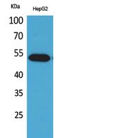
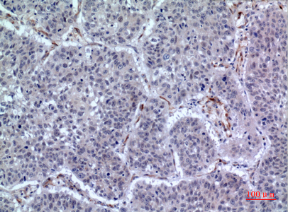
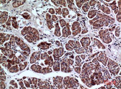
| WB, IHC-P |
|---|---|
| Primary Accession | Q8N149 |
| Reactivity | Human |
| Host | Rabbit |
| Clonality | Polyclonal |
| Calculated MW | 52966 Da |
| Gene ID | 11027 |
|---|---|
| Other Names | LILRA2; ILT1; LIR7; Leukocyte immunoglobulin-like receptor subfamily A member 2; CD85 antigen-like family member H; Immunoglobulin-like transcript 1; ILT-1; Leukocyte immunoglobulin-like receptor 7; LIR-7; CD85h |
| Dilution | WB~~Western Blot: 1/500 - 1/2000. IHC-p: 1/100-1/300. ELISA: 1/20000. Not yet tested in other applications. IHC-P~~N/A |
| Format | Liquid in PBS containing 50% glycerol, 0.5% BSA and 0.09% (W/V) sodium azide. |
| Storage Conditions | -20℃ |
| Name | LILRA2 |
|---|---|
| Synonyms | ILT1, LIR7 |
| Function | Part of the innate immune responses against microbial infection (PubMed:12529506, PubMed:27572839). Specifically recognizes a set of N-terminally truncated immunoglobulins that are produced via cleavage by proteases from a range of pathogenic bacteria and fungi, including L.pneumophila, M.hyorhinis, S.pneumoniae, S.aureus and C.albicans (PubMed:27572839). Recognizes epitopes that are in part in the variable region of the immunoglobulin light chains, but requires also the constant region for signaling (PubMed:27572839). Binds to a subset of cleaved IgM, IgG3 and IgG4 molecules, but does not bind cleaved IgA1 (PubMed:27572839). Binding of N-terminally truncated immunoglobulins mediates activation of neutrophils (PubMed:27572839). In monocytes, activation leads to the release of CSF2, CF3, IL6, CXCL8 and CCL3 and down-regulates responses to bacterial lipopolysaccharide (LPS), possibly via down-regulation of TLR4 expression and reduced signaling via TLR4 (PubMed:22479404). In eosinophils, activation by ligand binding leads to the release of RNASE2, IL4 and leukotriene C4 (PubMed:12529506). Does not bind class I MHC antigens (PubMed:19230061). |
| Cellular Location | Cell membrane; Single-pass type I membrane protein |
| Tissue Location | Detected on the surface of all peripheral blood monocytes, neutrophils, basophils and eosinophils (at protein level) (PubMed:12529506, PubMed:22479404). Expression levels are very low or not detectable on monocytes, T-cells, B-cells, dendritic cells and natural killer (NK) cells (PubMed:9548455) |

Thousands of laboratories across the world have published research that depended on the performance of antibodies from Abcepta to advance their research. Check out links to articles that cite our products in major peer-reviewed journals, organized by research category.
info@abcepta.com, and receive a free "I Love Antibodies" mug.

Provided below are standard protocols that you may find useful for product applications.
Background
Part of the innate immune responses against microbial infection (PubMed:12529506, PubMed:27572839). Specifically recognizes a set of N-terminally truncated immunoglobulins that are produced via cleavage by proteases from a range of pathogenic bacteria and fungi, including L.pneumophila, M.hyorhinis, S.pneumoniae, S.aureus and C.albicans (PubMed:27572839). Recognizes epitopes that are in part in the variable region of the immunoglobulin light chains, but requires also the constant region for signaling (PubMed:27572839). Binds to a subset of cleaved IgM, IgG3 and IgG4 molecules, but does not bind cleaved IgA1 (PubMed:27572839). Binding of N-terminally truncated immunoglobulins mediates activation of neutrophils (PubMed:27572839). In monocytes, activation leads to the release of CSF2, CF3, IL6, CXCL8 and CCL3 and down-regulates responses to bacterial lipopolysaccharide (LPS), possibly via down-regulation of TLR4 expression and reduced signaling via TLR4 (PubMed:22479404). In eosinophils, activation by ligand binding leads to the release of RNASE2, IL4 and leukotriene C4 (PubMed:12529506). Does not bind class I MHC antigens (PubMed:19230061).
If you have used an Abcepta product and would like to share how it has performed, please click on the "Submit Review" button and provide the requested information. Our staff will examine and post your review and contact you if needed.
If you have any additional inquiries please email technical services at tech@abcepta.com.
Ordering Information
Other Products
Shipping Information
