


 Foundational characteristics of cancer include proliferation, angiogenesis, migration, evasion of apoptosis, and cellular immortality. Find key markers for these cellular processes and antibodies to detect them.
 The SUMOplot™ Analysis Program predicts and scores sumoylation sites in your protein. SUMOylation is a post-translational modification involved in various cellular processes, such as nuclear-cytosolic transport, transcriptional regulation, apoptosis, protein stability, response to stress, and progression through the cell cycle.
 The Autophagy Receptor Motif Plotter predicts and scores autophagy receptor binding sites in your protein. Identifying proteins connected to this pathway is critical to understanding the role of autophagy in physiological as well as pathological processes such as development, differentiation, neurodegenerative diseases, stress, infection, and cancer.
ULK2 Polyclonal Antibody
- SPECIFICATION
- CITATIONS
- PROTOCOLS
- BACKGROUND

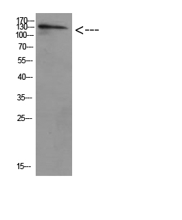
Application
| WB |
|---|---|
| Primary Accession | Q8IYT8 |
| Reactivity | Human, Mouse, Rat |
| Host | Rabbit |
| Clonality | Polyclonal |
| Calculated MW | 112694 Da |
| Gene ID | 9706 |
|---|---|
| Other Names | ULK2 KIAA0623 |
| Dilution | WB~~WB 1:500-2000, ELISA 1:10000-20000 |
| Format | Liquid in PBS containing 50% glycerol, 0.5% BSA and 0.09% (W/V) sodium azide. |
| Storage Conditions | -20℃ |
| Name | ULK2 |
|---|---|
| Synonyms | KIAA0623 |
| Function | Serine/threonine-protein kinase involved in autophagy in response to starvation. Acts upstream of phosphatidylinositol 3-kinase PIK3C3 to regulate the formation of autophagophores, the precursors of autophagosomes. Part of regulatory feedback loops in autophagy: acts both as a downstream effector and a negative regulator of mammalian target of rapamycin complex 1 (mTORC1) via interaction with RPTOR. Activated via phosphorylation by AMPK, also acts as a negative regulator of AMPK through phosphorylation of the AMPK subunits PRKAA1, PRKAB2 and PRKAG1. May phosphorylate ATG13/KIAA0652, FRS2, FRS3 and RPTOR; however such data need additional evidences. Not involved in ammonia-induced autophagy or in autophagic response of cerebellar granule neurons (CGN) to low potassium concentration. Plays a role early in neuronal differentiation and is required for granule cell axon formation: may govern axon formation via Ras-like GTPase signaling and through regulation of the Rab5-mediated endocytic pathways within developing axons. |
| Cellular Location | Cytoplasmic vesicle membrane; Peripheral membrane protein. Note=Localizes to pre-autophagosomal membrane |

Thousands of laboratories across the world have published research that depended on the performance of antibodies from Abcepta to advance their research. Check out links to articles that cite our products in major peer-reviewed journals, organized by research category.
info@abcepta.com, and receive a free "I Love Antibodies" mug.

Provided below are standard protocols that you may find useful for product applications.
Background
Serine/threonine-protein kinase involved in autophagy in response to starvation. Acts upstream of phosphatidylinositol 3- kinase PIK3C3 to regulate the formation of autophagophores, the precursors of autophagosomes. Part of regulatory feedback loops in autophagy: acts both as a downstream effector and a negative regulator of mammalian target of rapamycin complex 1 (mTORC1) via interaction with RPTOR. Activated via phosphorylation by AMPK, also acts as a negative regulator of AMPK through phosphorylation of the AMPK subunits PRKAA1, PRKAB2 and PRKAG1. May phosphorylate ATG13/KIAA0652, FRS2, FRS3 and RPTOR; however such data need additional evidences. Not involved in ammonia-induced autophagy or in autophagic response of cerebellar granule neurons (CGN) to low potassium concentration. Plays a role early in neuronal differentiation and is required for granule cell axon formation: may govern axon formation via Ras-like GTPase signaling and through regulation of the Rab5-mediated endocytic pathways within developing axons.
If you have used an Abcepta product and would like to share how it has performed, please click on the "Submit Review" button and provide the requested information. Our staff will examine and post your review and contact you if needed.
If you have any additional inquiries please email technical services at tech@abcepta.com.
Ordering Information
Other Products
Shipping Information
