


 Foundational characteristics of cancer include proliferation, angiogenesis, migration, evasion of apoptosis, and cellular immortality. Find key markers for these cellular processes and antibodies to detect them.
 The SUMOplot™ Analysis Program predicts and scores sumoylation sites in your protein. SUMOylation is a post-translational modification involved in various cellular processes, such as nuclear-cytosolic transport, transcriptional regulation, apoptosis, protein stability, response to stress, and progression through the cell cycle.
 The Autophagy Receptor Motif Plotter predicts and scores autophagy receptor binding sites in your protein. Identifying proteins connected to this pathway is critical to understanding the role of autophagy in physiological as well as pathological processes such as development, differentiation, neurodegenerative diseases, stress, infection, and cancer.
FA2H Polyclonal Antibody
- SPECIFICATION
- CITATIONS
- PROTOCOLS
- BACKGROUND

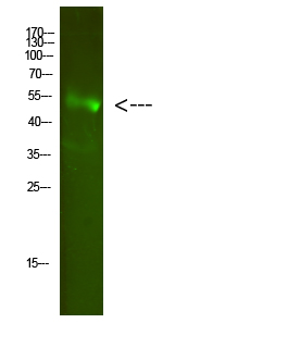
Application
| WB |
|---|---|
| Primary Accession | Q7L5A8 |
| Reactivity | Human, Mouse, Rat |
| Host | Rabbit |
| Clonality | Polyclonal |
| Calculated MW | 42791 Da |
| Gene ID | 79152 |
|---|---|
| Other Names | Fatty acid 2-hydroxylase (EC 1.-.-.-) (Fatty acid alpha-hydroxylase) |
| Dilution | WB~~WB 1:500-2000, ELISA 1:10000-20000 |
| Format | Liquid in PBS containing 50% glycerol, 0.5% BSA and 0.09% (W/V) sodium azide. |
| Storage Conditions | -20℃ |
| Name | FA2H |
|---|---|
| Function | Catalyzes the hydroxylation of free fatty acids at the C-2 position to produce 2-hydroxy fatty acids, which are building blocks of sphingolipids and glycosphingolipids common in neural tissue and epidermis (PubMed:15337768, PubMed:15863841, PubMed:17355976, PubMed:22517924). FA2H is stereospecific for the production of (R)-2- hydroxy fatty acids (PubMed:22517924). Plays an essential role in the synthesis of galactosphingolipids of the myelin sheath (By similarity). Responsible for the synthesis of sphingolipids and glycosphingolipids involved in the formation of epidermal lamellar bodies critical for skin permeability barrier (PubMed:17355976). Participates in the synthesis of glycosphingolipids and a fraction of type II wax diesters in sebaceous gland, specifically regulating hair follicle homeostasis (By similarity). Involved in the synthesis of sphingolipids of plasma membrane rafts, controlling lipid raft mobility and trafficking of raft-associated proteins (By similarity). |
| Cellular Location | Endoplasmic reticulum membrane {ECO:0000250|UniProtKB:Q5MPP0}; Multi-pass membrane protein. Microsome membrane; Multi-pass membrane protein |
| Tissue Location | Detected in differentiating cultured keratinocytes (at protein level). Detected in epidermis and cultured keratinocytes (PubMed:17355976). Highly expressed in brain and colon. Detected at lower levels in testis, prostate, pancreas and kidney (PubMed:15337768). |

Thousands of laboratories across the world have published research that depended on the performance of antibodies from Abcepta to advance their research. Check out links to articles that cite our products in major peer-reviewed journals, organized by research category.
info@abcepta.com, and receive a free "I Love Antibodies" mug.

Provided below are standard protocols that you may find useful for product applications.
Background
Catalyzes stereospecific hydroxylation of free fatty acids at the C-2 position to produce (R)-2-hydroxy fatty acids, which are building blocks of sphingolipids and glycosphingolipids common in neural tissue and epidermis (PubMed:17355976, PubMed:22517924, PubMed:15863841, PubMed:15337768). Plays an essential role in the synthesis of galactosphingolipids of the myelin sheath (By similarity). Responsible for the synthesis of sphingolipids and glycosphingolipids involved in the formation of epidermal lamellar bodies critical for skin permeability barrier (PubMed:17355976). Participates in the synthesis of glycosphingolipids and a fraction of type II wax diesters in sebaceous gland, specifically regulating hair follicle homeostasis (By similarity). Involved in the synthesis of sphingolipids of plasma membrane rafts, controlling lipid raft mobility and trafficking of raft-associated proteins (By similarity).
If you have used an Abcepta product and would like to share how it has performed, please click on the "Submit Review" button and provide the requested information. Our staff will examine and post your review and contact you if needed.
If you have any additional inquiries please email technical services at tech@abcepta.com.
Ordering Information
Other Products
Shipping Information
