


 Foundational characteristics of cancer include proliferation, angiogenesis, migration, evasion of apoptosis, and cellular immortality. Find key markers for these cellular processes and antibodies to detect them.
 The SUMOplot™ Analysis Program predicts and scores sumoylation sites in your protein. SUMOylation is a post-translational modification involved in various cellular processes, such as nuclear-cytosolic transport, transcriptional regulation, apoptosis, protein stability, response to stress, and progression through the cell cycle.
 The Autophagy Receptor Motif Plotter predicts and scores autophagy receptor binding sites in your protein. Identifying proteins connected to this pathway is critical to understanding the role of autophagy in physiological as well as pathological processes such as development, differentiation, neurodegenerative diseases, stress, infection, and cancer.
C1RL Polyclonal Antibody
- SPECIFICATION
- CITATIONS
- PROTOCOLS
- BACKGROUND

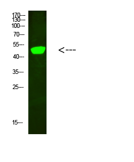
Application
| WB |
|---|---|
| Primary Accession | Q9NZP8 |
| Reactivity | Human |
| Host | Rabbit |
| Clonality | Polyclonal |
| Calculated MW | 53498 Da |
| Gene ID | 51279 |
|---|---|
| Other Names | Complement C1r subcomponent-like protein (C1r-LP) (C1r-like protein) (EC 3.4.21.-) (C1r-like serine protease analog protein) (CLSPa) |
| Dilution | WB~~WB 1:500-2000, ELISA 1:10000-20000 |
| Format | Liquid in PBS containing 50% glycerol, 0.5% BSA and 0.09% (W/V) sodium azide. |
| Storage Conditions | -20℃ |
| Name | C1RL |
|---|---|
| Synonyms | C1RL1, C1RLP, CLSPA |
| Function | Mediates the proteolytic cleavage of HP/haptoglobin in the endoplasmic reticulum. |
| Cellular Location | Secreted. |
| Tissue Location | Highly expressed in placenta, liver, kidney, pancreas, moderately in lung, spleen, prostate, ovary, colon, and PBL, and weakly in heart, skeletal muscle, thymus, testis, and small intestine. Expressed in PC-3 (prostate adenocarcinoma) and SK-OV-3 (ovary adenocarcinoma) cells, but not in LoVo and HT-29 (colon adenocarcinoma), SMMC7721 (hepatocellular carcinoma), CaoV-3 (ovary adenocarcinoma), HeLa (cervix epithelioid carcinoma), MCF-7 (breast adenocarcinoma), U-251MG (glioma) or A-549 (lung carcinoma) cells Widely expressed in myeloid leukemia cell lines, including K-562 (chronic myelogenous leukemia), THP-1 (myelomonocytic leukemia), HL-60 and NB4 (promyelocytic leukemia), and KG-1 (acute myelogenous leukemia) cells. Expressed mainly in the liver and in serum (at protein level) |

Thousands of laboratories across the world have published research that depended on the performance of antibodies from Abcepta to advance their research. Check out links to articles that cite our products in major peer-reviewed journals, organized by research category.
info@abcepta.com, and receive a free "I Love Antibodies" mug.

Provided below are standard protocols that you may find useful for product applications.
Background
Mediates the proteolytic cleavage of HP/haptoglobin in the endoplasmic reticulum.
If you have used an Abcepta product and would like to share how it has performed, please click on the "Submit Review" button and provide the requested information. Our staff will examine and post your review and contact you if needed.
If you have any additional inquiries please email technical services at tech@abcepta.com.
Ordering Information
Shipping Information
