


 Foundational characteristics of cancer include proliferation, angiogenesis, migration, evasion of apoptosis, and cellular immortality. Find key markers for these cellular processes and antibodies to detect them.
 The SUMOplot™ Analysis Program predicts and scores sumoylation sites in your protein. SUMOylation is a post-translational modification involved in various cellular processes, such as nuclear-cytosolic transport, transcriptional regulation, apoptosis, protein stability, response to stress, and progression through the cell cycle.
 The Autophagy Receptor Motif Plotter predicts and scores autophagy receptor binding sites in your protein. Identifying proteins connected to this pathway is critical to understanding the role of autophagy in physiological as well as pathological processes such as development, differentiation, neurodegenerative diseases, stress, infection, and cancer.
FGFR1 Antibody (N-term)
Purified Rabbit Polyclonal Antibody (Pab)
- SPECIFICATION
- CITATIONS: 9
- PROTOCOLS
- BACKGROUND

Application
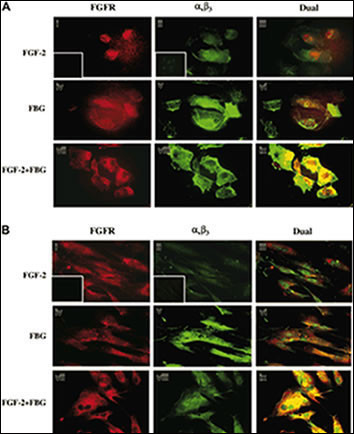
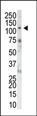
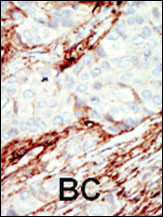
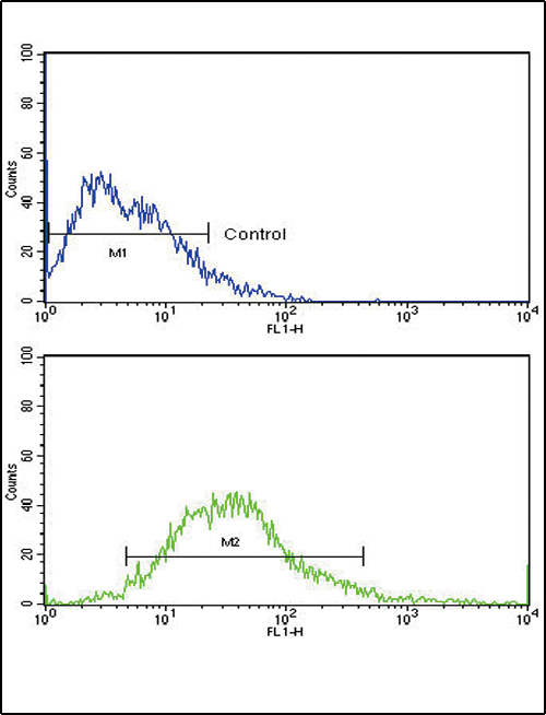
| IF, IHC-P, WB, FC, E |
|---|---|
| Primary Accession | P11362 |
| Reactivity | Human, Mouse |
| Host | Rabbit |
| Clonality | Polyclonal |
| Isotype | Rabbit IgG |
| Calculated MW | 91868 Da |
| Antigen Region | 19-48 aa |
| Gene ID | 2260 |
|---|---|
| Other Names | Fibroblast growth factor receptor 1, FGFR-1, Basic fibroblast growth factor receptor 1, BFGFR, bFGF-R-1, Fms-like tyrosine kinase 2, FLT-2, N-sam, Proto-oncogene c-Fgr, CD331, FGFR1, BFGFR, CEK, FGFBR, FLG, FLT2, HBGFR |
| Target/Specificity | This FGFR1 antibody is generated from rabbits immunized with a KLH conjugated synthetic peptide between 19~48 amino acids from the N-terminal region of human FGFR1. |
| Dilution | IF~~1:50~100 IHC-P~~1:50~100 WB~~1:1000 FC~~1:10~50 E~~Use at an assay dependent concentration. |
| Format | Purified polyclonal antibody supplied in PBS with 0.09% (W/V) sodium azide. This antibody is prepared by Saturated Ammonium Sulfate (SAS) precipitation followed by dialysis against PBS. |
| Storage | Maintain refrigerated at 2-8°C for up to 2 weeks. For long term storage store at -20°C in small aliquots to prevent freeze-thaw cycles. |
| Precautions | FGFR1 Antibody (N-term) is for research use only and not for use in diagnostic or therapeutic procedures. |
| Name | FGFR1 |
|---|---|
| Synonyms | BFGFR, CEK, FGFBR, FLG, FLT2, HBGFR |
| Function | Tyrosine-protein kinase that acts as a cell-surface receptor for fibroblast growth factors and plays an essential role in the regulation of embryonic development, cell proliferation, differentiation and migration. Required for normal mesoderm patterning and correct axial organization during embryonic development, normal skeletogenesis and normal development of the gonadotropin-releasing hormone (GnRH) neuronal system. Phosphorylates PLCG1, FRS2, GAB1 and SHB. Ligand binding leads to the activation of several signaling cascades. Activation of PLCG1 leads to the production of the cellular signaling molecules diacylglycerol and inositol 1,4,5-trisphosphate. Phosphorylation of FRS2 triggers recruitment of GRB2, GAB1, PIK3R1 and SOS1, and mediates activation of RAS, MAPK1/ERK2, MAPK3/ERK1 and the MAP kinase signaling pathway, as well as of the AKT1 signaling pathway. Promotes phosphorylation of SHC1, STAT1 and PTPN11/SHP2. In the nucleus, enhances RPS6KA1 and CREB1 activity and contributes to the regulation of transcription. FGFR1 signaling is down-regulated by IL17RD/SEF, and by FGFR1 ubiquitination, internalization and degradation. |
| Cellular Location | Cell membrane; Single-pass type I membrane protein. Nucleus. Cytoplasm, cytosol. Cytoplasmic vesicle. Note=After ligand binding, both receptor and ligand are rapidly internalized. Can translocate to the nucleus after internalization, or by translocation from the endoplasmic reticulum or Golgi apparatus to the cytosol, and from there to the nucleus |
| Tissue Location | Detected in astrocytoma, neuroblastoma and adrenal cortex cell lines. Some isoforms are detected in foreskin fibroblast cell lines, however isoform 17, isoform 18 and isoform 19 are not detected in these cells. |

Provided below are standard protocols that you may find useful for product applications.
Background
FGFR1 is a member of the fibroblast growth factor receptor family, where amino acid sequence is highly conserved between members and throughout evolution. FGFR family members differ from one another in their ligand affinities and tissue distribution. A full-length representative protein consists of an extracellular region, composed of three immunoglobulin-like domains, a single hydrophobic membrane-spanning segment and a cytoplasmic tyrosine kinase domain. The extracellular portion of the protein interacts with fibroblast growth factors, setting in motion a cascade of downstream signals, ultimately influencing mitogenesis and differentiation. This particular family member binds both acidic and basic fibroblast growth factors and is involved in limb induction. Mutations in this gene can lead to Pfeiffer syndrome and Jackson-Weiss syndrome. The genomic organization of the gene is very similar to family members 2-4, encompassing 19 exons that are subject to complex alternative splicing, which allows for structural, tissue expression and ligand affinity variations among the isoforms.
References
Jiao, J., et al., Arch. Biochem. Biophys. 410(2):187-200 (2003).
Fu, L., et al., J. Comp. Neurol. 462(2):265-273 (2003).
Lundin, L., et al., Exp. Cell Res. 287(1):190-198 (2003).
Kiselyov, V.V., et al., Structure (Camb.) 11(6):691-701 (2003).
Baumann, H., et al., J. Biol. Chem. 278(18):16198-16208 (2003).
If you have used an Abcepta product and would like to share how it has performed, please click on the "Submit Review" button and provide the requested information. Our staff will examine and post your review and contact you if needed.
If you have any additional inquiries please email technical services at tech@abcepta.com.
Ordering Information
Other Products
Shipping Information
