


 Foundational characteristics of cancer include proliferation, angiogenesis, migration, evasion of apoptosis, and cellular immortality. Find key markers for these cellular processes and antibodies to detect them.
 The SUMOplot™ Analysis Program predicts and scores sumoylation sites in your protein. SUMOylation is a post-translational modification involved in various cellular processes, such as nuclear-cytosolic transport, transcriptional regulation, apoptosis, protein stability, response to stress, and progression through the cell cycle.
 The Autophagy Receptor Motif Plotter predicts and scores autophagy receptor binding sites in your protein. Identifying proteins connected to this pathway is critical to understanding the role of autophagy in physiological as well as pathological processes such as development, differentiation, neurodegenerative diseases, stress, infection, and cancer.
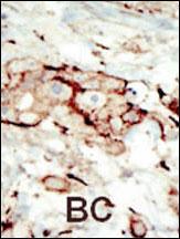
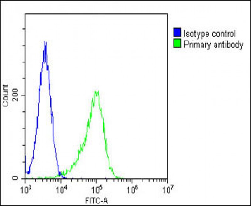
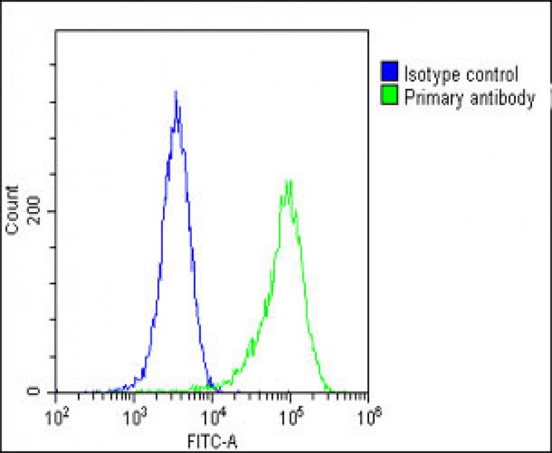
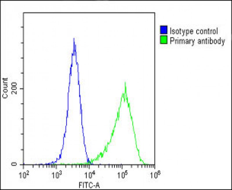
FGFR2 Antibody (N-term)
Affinity Purified Rabbit Polyclonal Antibody (Pab)
- SPECIFICATION
- CITATIONS: 6
- PROTOCOLS
- BACKGROUND

Application
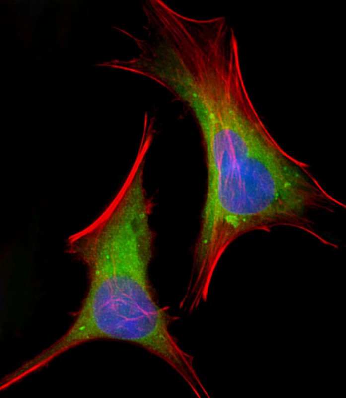
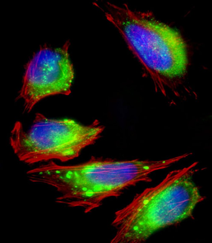
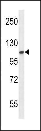
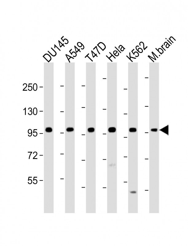
| IHC-P, WB, FC, IF, E |
|---|---|
| Primary Accession | P21802 |
| Other Accession | P21803 |
| Reactivity | Human, Mouse |
| Host | Rabbit |
| Clonality | Polyclonal |
| Isotype | Rabbit IgG |
| Calculated MW | 92025 Da |
| Antigen Region | 22-51 aa |
| Gene ID | 2263 |
|---|---|
| Other Names | Fibroblast growth factor receptor 2, FGFR-2, K-sam, KGFR, Keratinocyte growth factor receptor, CD332, FGFR2, BEK, KGFR, KSAM |
| Target/Specificity | This FGFR2 antibody is generated from rabbits immunized with a KLH conjugated synthetic peptide between 22-51 amino acids from the N-terminal region of human FGFR2. |
| Dilution | IHC-P~~1:50~100 WB~~1:1000-1:2000 FC~~1:25 IF~~1:25 E~~Use at an assay dependent concentration. |
| Format | Purified polyclonal antibody supplied in PBS with 0.09% (W/V) sodium azide. This antibody is purified through a protein A column, followed by peptide affinity purification. |
| Storage | Maintain refrigerated at 2-8°C for up to 2 weeks. For long term storage store at -20°C in small aliquots to prevent freeze-thaw cycles. |
| Precautions | FGFR2 Antibody (N-term) is for research use only and not for use in diagnostic or therapeutic procedures. |
| Name | FGFR2 |
|---|---|
| Synonyms | BEK, KGFR, KSAM |
| Function | Tyrosine-protein kinase that acts as a cell-surface receptor for fibroblast growth factors and plays an essential role in the regulation of cell proliferation, differentiation, migration and apoptosis, and in the regulation of embryonic development. Required for normal embryonic patterning, trophoblast function, limb bud development, lung morphogenesis, osteogenesis and skin development. Plays an essential role in the regulation of osteoblast differentiation, proliferation and apoptosis, and is required for normal skeleton development. Promotes cell proliferation in keratinocytes and immature osteoblasts, but promotes apoptosis in differentiated osteoblasts. Phosphorylates PLCG1, FRS2 and PAK4. Ligand binding leads to the activation of several signaling cascades. Activation of PLCG1 leads to the production of the cellular signaling molecules diacylglycerol and inositol 1,4,5-trisphosphate. Phosphorylation of FRS2 triggers recruitment of GRB2, GAB1, PIK3R1 and SOS1, and mediates activation of RAS, MAPK1/ERK2, MAPK3/ERK1 and the MAP kinase signaling pathway, as well as of the AKT1 signaling pathway. FGFR2 signaling is down-regulated by ubiquitination, internalization and degradation. Mutations that lead to constitutive kinase activation or impair normal FGFR2 maturation, internalization and degradation lead to aberrant signaling. Over-expressed FGFR2 promotes activation of STAT1. |
| Cellular Location | Cell membrane; Single-pass type I membrane protein. Golgi apparatus. Cytoplasmic vesicle. Note=Detected on osteoblast plasma membrane lipid rafts. After ligand binding, the activated receptor is rapidly internalized and degraded [Isoform 3]: Cell membrane; Single-pass type I membrane protein. Note=After ligand binding, the activated receptor is rapidly internalized and degraded [Isoform 13]: Secreted. |

Provided below are standard protocols that you may find useful for product applications.
Background
FGFR2 is a member of the fibroblast growth factor receptor family, where amino acid sequence is highly conserved between members and throughout evolution. FGFR family members differ from one another in their ligand affinities and tissue distribution. A full-length representative protein consists of an extracellular region, composed of three immunoglobulin-like domains, a single hydrophobic membrane-spanning segment and a cytoplasmic tyrosine kinase domain. The extracellular portion of the protein interacts with fibroblast growth factors, setting in motion a cascade of downstream signals, ultimately influencing mitogenesis and differentiation. This particular family member is a high-affinity receptor for acidic, basic and/or keratinocyte growth factor, depending on the isoform. Mutations in the gene are associated with many craniosynostotic syndromes and bone malformations. The genomic organization of the gene encompasses 20 exons. Alternative splicing in multiple exons, including those encoding the Ig-like domains, the transmembrane region and the carboxyl terminus, results in varied isoforms which differ in structure and specificity. Isoform 1 has equal affinity for aFGF and bFGF but does not bind KGF.
References
Freeman, K.W., et al., Cancer Res. 63(19):6237-6243 (2003). Goriely, A., et al., Science 301(5633):643-646 (2003). Fomenkov, A., et al., J. Biol. Chem. 278(26):23906-23914 (2003). Katoh, M., et al., Int. J. Mol. Med. 11(5):579-583 (2003). Katoh, M., et al., Int. J. Oncol. 22(5):1155-1159 (2003).
If you have used an Abcepta product and would like to share how it has performed, please click on the "Submit Review" button and provide the requested information. Our staff will examine and post your review and contact you if needed.
If you have any additional inquiries please email technical services at tech@abcepta.com.
Ordering Information
Other Products
Shipping Information
