


 Foundational characteristics of cancer include proliferation, angiogenesis, migration, evasion of apoptosis, and cellular immortality. Find key markers for these cellular processes and antibodies to detect them.
 The SUMOplot™ Analysis Program predicts and scores sumoylation sites in your protein. SUMOylation is a post-translational modification involved in various cellular processes, such as nuclear-cytosolic transport, transcriptional regulation, apoptosis, protein stability, response to stress, and progression through the cell cycle.
 The Autophagy Receptor Motif Plotter predicts and scores autophagy receptor binding sites in your protein. Identifying proteins connected to this pathway is critical to understanding the role of autophagy in physiological as well as pathological processes such as development, differentiation, neurodegenerative diseases, stress, infection, and cancer.
CCK4 (PTK7) Antibody (N-term)
Purified Rabbit Polyclonal Antibody (Pab)
- SPECIFICATION
- CITATIONS: 1
- PROTOCOLS
- BACKGROUND

Application
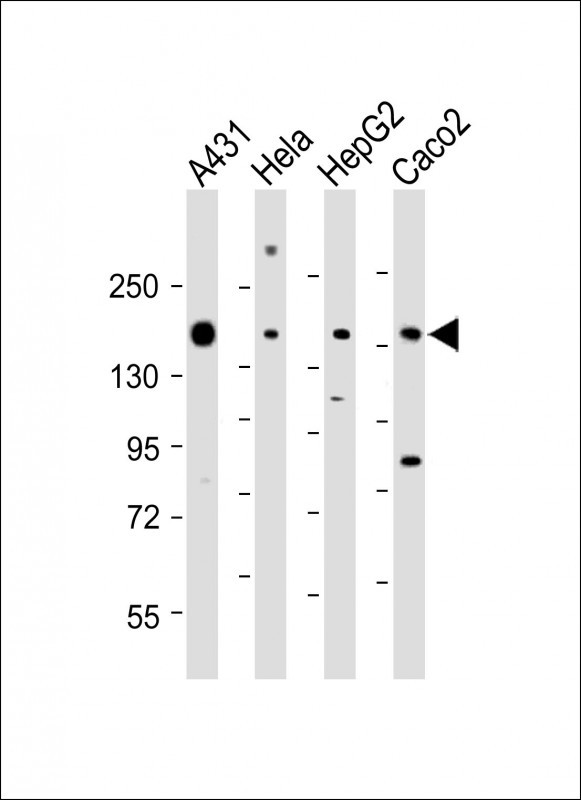
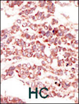
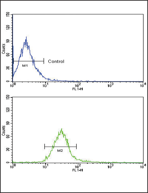
| IHC-P, FC, WB, E |
|---|---|
| Primary Accession | Q13308 |
| Reactivity | Human |
| Host | Rabbit |
| Clonality | Polyclonal |
| Isotype | Rabbit IgG |
| Calculated MW | 118392 Da |
| Antigen Region | 21-52 aa |
| Gene ID | 5754 |
|---|---|
| Other Names | Inactive tyrosine-protein kinase 7, Colon carcinoma kinase 4, CCK-4, Protein-tyrosine kinase 7, Pseudo tyrosine kinase receptor 7, Tyrosine-protein kinase-like 7, PTK7, CCK4 |
| Target/Specificity | This CCK4 (PTK7) antibody is generated from rabbits immunized with a KLH conjugated synthetic peptide between 21-52 amino acids from the N-terminal region of human CCK4 (PTK7). |
| Dilution | IHC-P~~1:50~100 FC~~1:10~50 WB~~1:1000-1:2000 E~~Use at an assay dependent concentration. |
| Format | Purified polyclonal antibody supplied in PBS with 0.09% (W/V) sodium azide. This antibody is purified through a protein A column, followed by peptide affinity purification. |
| Storage | Maintain refrigerated at 2-8°C for up to 2 weeks. For long term storage store at -20°C in small aliquots to prevent freeze-thaw cycles. |
| Precautions | CCK4 (PTK7) Antibody (N-term) is for research use only and not for use in diagnostic or therapeutic procedures. |
| Name | PTK7 |
|---|---|
| Synonyms | CCK4 |
| Function | Inactive tyrosine kinase involved in Wnt signaling pathway. Component of both the non-canonical (also known as the Wnt/planar cell polarity signaling) and the canonical Wnt signaling pathway. Functions in cell adhesion, cell migration, cell polarity, proliferation, actin cytoskeleton reorganization and apoptosis. Has a role in embryogenesis, epithelial tissue organization and angiogenesis. |
| Cellular Location | Membrane; Single- pass type I membrane protein. Cell junction. Note=Colocalizes with MMP14 at cell junctions. Also localizes at the leading edge of migrating cells |
| Tissue Location | Highly expressed in lung, liver, pancreas, kidney, placenta and melanocytes. Weakly expressed in thyroid gland, ovary, brain, heart and skeletal muscle. Also expressed in erythroleukemia cells. But not expressed in colon |

Provided below are standard protocols that you may find useful for product applications.
Background
CCK4 may function as a cell adhesion molecule. Although it belongs to the insuline receptor subfamily of the Tyr protein kinases, it likely lacks the catalytic activity of a tyrosine kinase. It may be connected to the pathophysiology of colon carcinomas and/or may represent a tumor progression marker. This Type I membrane protein is highly expressed in lung, liver, pancreas, kidney, placenta and melanocytes, but weakly expressed in thyroid gland, ovary, brain, heart and skeletal muscle, and not in colon. It is also expressed in erythroleukemia cells.
References
Zhang, H., et al., Nat. Biotechnol. 21(6):660-666 (2003).
Park, S.K., et al., J. Biochem. 119(2):235-239 (1996).
Mossie, K., et al., Oncogene 11(10):2179-2184 (1995).
If you have used an Abcepta product and would like to share how it has performed, please click on the "Submit Review" button and provide the requested information. Our staff will examine and post your review and contact you if needed.
If you have any additional inquiries please email technical services at tech@abcepta.com.
Ordering Information
Other Products
Shipping Information
