


 Foundational characteristics of cancer include proliferation, angiogenesis, migration, evasion of apoptosis, and cellular immortality. Find key markers for these cellular processes and antibodies to detect them.
 The SUMOplot™ Analysis Program predicts and scores sumoylation sites in your protein. SUMOylation is a post-translational modification involved in various cellular processes, such as nuclear-cytosolic transport, transcriptional regulation, apoptosis, protein stability, response to stress, and progression through the cell cycle.
 The Autophagy Receptor Motif Plotter predicts and scores autophagy receptor binding sites in your protein. Identifying proteins connected to this pathway is critical to understanding the role of autophagy in physiological as well as pathological processes such as development, differentiation, neurodegenerative diseases, stress, infection, and cancer.
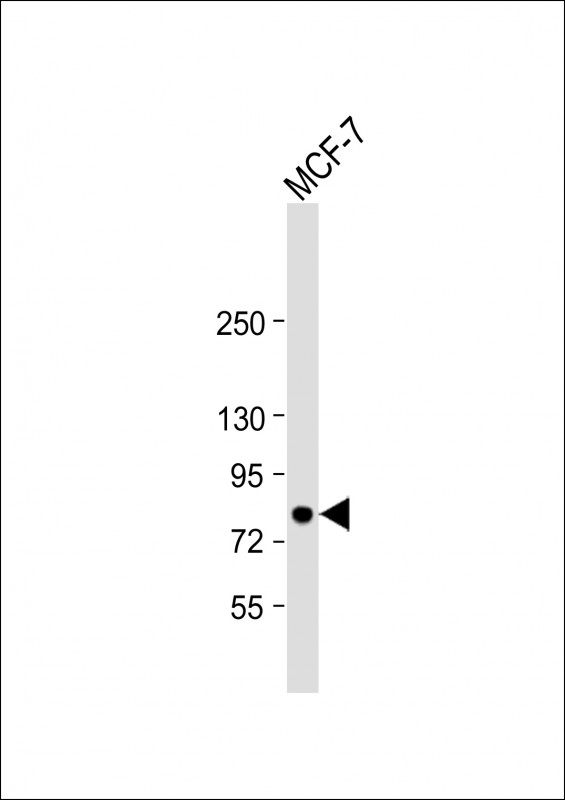
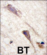
B-RAF Antibody (S601)
Affinity Purified Rabbit Polyclonal Antibody (Pab)
- SPECIFICATION
- CITATIONS
- PROTOCOLS
- BACKGROUND

Application
| IHC-P, WB, E |
|---|---|
| Primary Accession | P15056 |
| Other Accession | P28028, Q04982 |
| Reactivity | Human |
| Predicted | Chicken, Mouse |
| Host | Rabbit |
| Clonality | Polyclonal |
| Isotype | Rabbit IgG |
| Calculated MW | 84437 Da |
| Antigen Region | 580-609 aa |
| Gene ID | 673 |
|---|---|
| Other Names | Serine/threonine-protein kinase B-raf, Proto-oncogene B-Raf, p94, v-Raf murine sarcoma viral oncogene homolog B1, BRAF, BRAF1, RAFB1 |
| Target/Specificity | This B-RAF antibody is generated from rabbits immunized with a KLH conjugated synthetic peptide between 580-609 amino acids from human B-RAF. |
| Dilution | IHC-P~~1:10~50 WB~~1:1000 E~~Use at an assay dependent concentration. |
| Format | Purified polyclonal antibody supplied in PBS with 0.09% (W/V) sodium azide. This antibody is purified through a protein A column, followed by peptide affinity purification. |
| Storage | Maintain refrigerated at 2-8°C for up to 2 weeks. For long term storage store at -20°C in small aliquots to prevent freeze-thaw cycles. |
| Precautions | B-RAF Antibody (S601) is for research use only and not for use in diagnostic or therapeutic procedures. |
| Name | BRAF (HGNC:1097) |
|---|---|
| Synonyms | BRAF1, RAFB1 |
| Function | Protein kinase involved in the transduction of mitogenic signals from the cell membrane to the nucleus (Probable). Phosphorylates MAP2K1, and thereby activates the MAP kinase signal transduction pathway (PubMed:21441910, PubMed:29433126). Phosphorylates PFKFB2 (PubMed:36402789). May play a role in the postsynaptic responses of hippocampal neurons (PubMed:1508179). |
| Cellular Location | Nucleus. Cytoplasm. Cell membrane. Note=Colocalizes with RGS14 and RAF1 in both the cytoplasm and membranes. |
| Tissue Location | Brain and testis. |

Thousands of laboratories across the world have published research that depended on the performance of antibodies from Abcepta to advance their research. Check out links to articles that cite our products in major peer-reviewed journals, organized by research category.
info@abcepta.com, and receive a free "I Love Antibodies" mug.

Provided below are standard protocols that you may find useful for product applications.
Background
BRAF, a member of the RAF subfamily of Ser/Thr protein kinases, is involved in the transduction of mitogenic signals from the cell membrane to the nucleus. It may play a role in the postsynaptic responses of hippocampal neurons. This cytoplasmic protein is expressed in brain and testis. Defects in BRAF are involved in a wide range of cancers including lung cancer and non-Hodgkin lymphoma (NHL). This protein contains 1 zinc-dependent phorbol-ester and DAG binding domain.
References
Hingorani, S.R., et al., Cancer Res. 63(17):5198-5202 (2003).
Lee, J.W., et al., Br. J. Cancer 89(10):1958-1960 (2003).
Davies, H., et al., Nature 417(6892):949-954 (2002).
Naoki, K., et al., Cancer Res. 62(23):7001-7003 (2002).
Stephens, R.M., et al., Mol. Cell. Biol. 12(9):3733-3742 (1992).
If you have used an Abcepta product and would like to share how it has performed, please click on the "Submit Review" button and provide the requested information. Our staff will examine and post your review and contact you if needed.
If you have any additional inquiries please email technical services at tech@abcepta.com.
Ordering Information
Other Products
Shipping Information
