


 Foundational characteristics of cancer include proliferation, angiogenesis, migration, evasion of apoptosis, and cellular immortality. Find key markers for these cellular processes and antibodies to detect them.
 The SUMOplot™ Analysis Program predicts and scores sumoylation sites in your protein. SUMOylation is a post-translational modification involved in various cellular processes, such as nuclear-cytosolic transport, transcriptional regulation, apoptosis, protein stability, response to stress, and progression through the cell cycle.
 The Autophagy Receptor Motif Plotter predicts and scores autophagy receptor binding sites in your protein. Identifying proteins connected to this pathway is critical to understanding the role of autophagy in physiological as well as pathological processes such as development, differentiation, neurodegenerative diseases, stress, infection, and cancer.
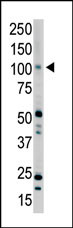
PRK2 Antibody (C-term)
Purified Rabbit Polyclonal Antibody (Pab)
- SPECIFICATION
- CITATIONS
- PROTOCOLS
- BACKGROUND

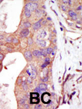
Application
| WB, IHC-P, E |
|---|---|
| Primary Accession | Q16513 |
| Reactivity | Human |
| Host | Rabbit |
| Clonality | Polyclonal |
| Isotype | Rabbit IgG |
| Calculated MW | 112035 Da |
| Antigen Region | 627-658 aa |
| Gene ID | 5586 |
|---|---|
| Other Names | Serine/threonine-protein kinase N2, PKN gamma, Protein kinase C-like 2, Protein-kinase C-related kinase 2, PKN2, PRK2, PRKCL2 |
| Target/Specificity | This PRK2 antibody is generated from rabbits immunized with a KLH conjugated synthetic peptide between 627-658 amino acids from the C-terminal region of human PRK2. |
| Dilution | WB~~1:1000 IHC-P~~1:50~100 E~~Use at an assay dependent concentration. |
| Format | Purified polyclonal antibody supplied in PBS with 0.09% (W/V) sodium azide. This antibody is prepared by Saturated Ammonium Sulfate (SAS) precipitation followed by dialysis against PBS. |
| Storage | Maintain refrigerated at 2-8°C for up to 2 weeks. For long term storage store at -20°C in small aliquots to prevent freeze-thaw cycles. |
| Precautions | PRK2 Antibody (C-term) is for research use only and not for use in diagnostic or therapeutic procedures. |
| Name | PKN2 |
|---|---|
| Synonyms | PRK2, PRKCL2 |
| Function | PKC-related serine/threonine-protein kinase and Rho/Rac effector protein that participates in specific signal transduction responses in the cell. Plays a role in the regulation of cell cycle progression, actin cytoskeleton assembly, cell migration, cell adhesion, tumor cell invasion and transcription activation signaling processes. Phosphorylates CTTN in hyaluronan-induced astrocytes and hence decreases CTTN ability to associate with filamentous actin. Phosphorylates HDAC5, therefore lead to impair HDAC5 import. Direct RhoA target required for the regulation of the maturation of primordial junctions into apical junction formation in bronchial epithelial cells. Required for G2/M phases of the cell cycle progression and abscission during cytokinesis in a ECT2-dependent manner. Stimulates FYN kinase activity that is required for establishment of skin cell-cell adhesion during keratinocytes differentiation. Regulates epithelial bladder cells speed and direction of movement during cell migration and tumor cell invasion. Inhibits Akt pro-survival-induced kinase activity. Mediates Rho protein-induced transcriptional activation via the c-fos serum response factor (SRF). Involved in the negative regulation of ciliogenesis (PubMed:27104747). |
| Cellular Location | Cytoplasm. Nucleus Membrane {ECO:0000250|UniProtKB:Q8BWW9}. Cell projection, lamellipodium. Cytoplasm, cytoskeleton. Cleavage furrow. Midbody Cell junction. Note=Colocalizes with PTPN13 in lamellipodia-like structures, regions of large actin turnover. Accumulates during telophase at the cleavage furrow and concentrates finally around the midbody in cytokinesis. Recruited to nascent cell-cell contacts at the apical surface of cells. In the course of viral infection, colocalizes with HCV NS5B at perinuclear region in the cytoplasm. |
| Tissue Location | Ubiquitous. Expressed in numerous tumor cell lines, especially in bladder tumor cells. |

Thousands of laboratories across the world have published research that depended on the performance of antibodies from Abcepta to advance their research. Check out links to articles that cite our products in major peer-reviewed journals, organized by research category.
info@abcepta.com, and receive a free "I Love Antibodies" mug.

Provided below are standard protocols that you may find useful for product applications.
Background
Protein kinases are enzymes that transfer a phosphate group from a phosphate donor, generally the g phosphate of ATP, onto an acceptor amino acid in a substrate protein. By this basic mechanism, protein kinases mediate most of the signal transduction in eukaryotic cells, regulating cellular metabolism, transcription, cell cycle progression, cytoskeletal rearrangement and cell movement, apoptosis, and differentiation. With more than 500 gene products, the protein kinase family is one of the largest families of proteins in eukaryotes. The family has been classified in 8 major groups based on sequence comparison of their tyrosine (PTK) or serine/threonine (STK) kinase catalytic domains. The AGC kinase group consists of 63 kinases including the cyclic nucleotide-regulated protein kinase (PKA & PKG) family, the diacylglycerol-activated/phospholipid-dependent protein kinase C (PKC) family, the related to PKA and PKC (RAC/Akt) protein kinase family, the kinases that phosphorylate G protein-coupled receptors family (ARK), and the kinases that phosphorylate ribosomal protein S6 family (RSK). The calcium/calmodulin-dependent kinase (CAMK) group consists of 75 kinases regulated by Ca2+/CaM and close relative family (CAMK, CAMKL, DAPK, MAPKAPK).
References
Yu, W., et al., J. Biol. Chem. 272(15):10030-10034 (1997).
Palmer, R.H., et al., FEBS Lett. 356(1):5-8 (1994).
Palmer, R.H., et al., Eur. J. Biochem. 227 (1-2), 344-351 (1995).
If you have used an Abcepta product and would like to share how it has performed, please click on the "Submit Review" button and provide the requested information. Our staff will examine and post your review and contact you if needed.
If you have any additional inquiries please email technical services at tech@abcepta.com.
Ordering Information
Other Products
Shipping Information
