


 Foundational characteristics of cancer include proliferation, angiogenesis, migration, evasion of apoptosis, and cellular immortality. Find key markers for these cellular processes and antibodies to detect them.
 The SUMOplot™ Analysis Program predicts and scores sumoylation sites in your protein. SUMOylation is a post-translational modification involved in various cellular processes, such as nuclear-cytosolic transport, transcriptional regulation, apoptosis, protein stability, response to stress, and progression through the cell cycle.
 The Autophagy Receptor Motif Plotter predicts and scores autophagy receptor binding sites in your protein. Identifying proteins connected to this pathway is critical to understanding the role of autophagy in physiological as well as pathological processes such as development, differentiation, neurodegenerative diseases, stress, infection, and cancer.
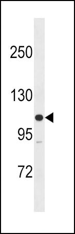
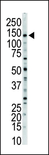
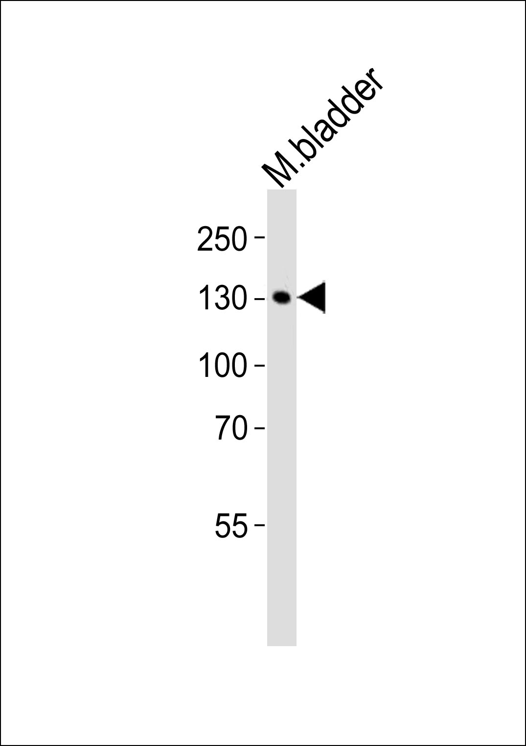
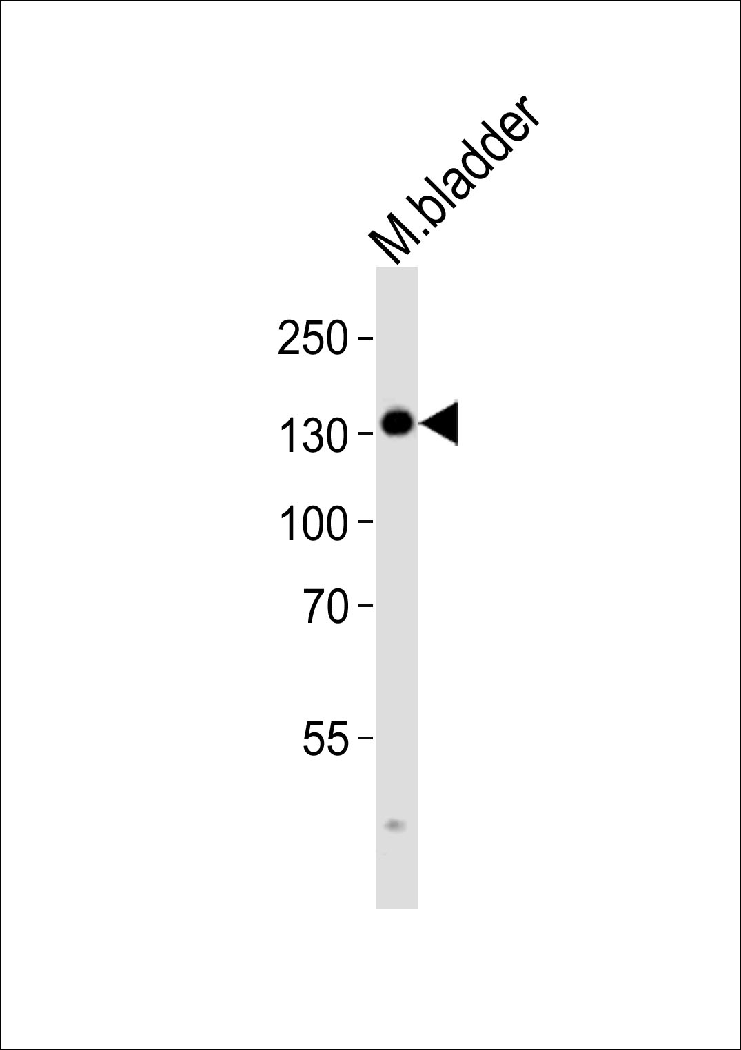
MLCK Antibody (N-term)
Purified Rabbit Polyclonal Antibody (Pab)
- SPECIFICATION
- CITATIONS: 3
- PROTOCOLS
- BACKGROUND

Application
| WB, IHC-P, E |
|---|---|
| Primary Accession | Q15746 |
| Other Accession | P29294 |
| Reactivity | Human, Mouse |
| Predicted | Rabbit |
| Host | Rabbit |
| Clonality | Polyclonal |
| Isotype | Rabbit IgG |
| Calculated MW | 210715 Da |
| Antigen Region | 923-953 aa |
| Gene ID | 4638 |
|---|---|
| Other Names | Myosin light chain kinase, smooth muscle, MLCK, smMLCK, Kinase-related protein, KRP, Telokin, Myosin light chain kinase, smooth muscle, deglutamylated form, MYLK, MLCK, MLCK1, MYLK1 |
| Target/Specificity | This MLCK antibody is generated from rabbits immunized with a KLH conjugated synthetic peptide between 923-953 amino acids from the N-terminal region of human MLCK. |
| Dilution | WB~~1:1000 IHC-P~~N/A E~~Use at an assay dependent concentration. |
| Format | Purified polyclonal antibody supplied in PBS with 0.09% (W/V) sodium azide. This antibody is prepared by Saturated Ammonium Sulfate (SAS) precipitation followed by dialysis against PBS. |
| Storage | Maintain refrigerated at 2-8°C for up to 2 weeks. For long term storage store at -20°C in small aliquots to prevent freeze-thaw cycles. |
| Precautions | MLCK Antibody (N-term) is for research use only and not for use in diagnostic or therapeutic procedures. |
| Name | MYLK (HGNC:7590) |
|---|---|
| Synonyms | MLCK, MLCK1, MYLK1 |
| Function | Calcium/calmodulin-dependent myosin light chain kinase implicated in smooth muscle contraction via phosphorylation of myosin light chains (MLC). Also regulates actin-myosin interaction through a non-kinase activity. Phosphorylates PTK2B/PYK2 and myosin light-chains. Involved in the inflammatory response (e.g. apoptosis, vascular permeability, leukocyte diapedesis), cell motility and morphology, airway hyperreactivity and other activities relevant to asthma. Required for tonic airway smooth muscle contraction that is necessary for physiological and asthmatic airway resistance. Necessary for gastrointestinal motility. Implicated in the regulation of endothelial as well as vascular permeability, probably via the regulation of cytoskeletal rearrangements. In the nervous system it has been shown to control the growth initiation of astrocytic processes in culture and to participate in transmitter release at synapses formed between cultured sympathetic ganglion cells. Critical participant in signaling sequences that result in fibroblast apoptosis. Plays a role in the regulation of epithelial cell survival. Required for epithelial wound healing, especially during actomyosin ring contraction during purse-string wound closure. Mediates RhoA-dependent membrane blebbing. Triggers TRPC5 channel activity in a calcium-dependent signaling, by inducing its subcellular localization at the plasma membrane. Promotes cell migration (including tumor cells) and tumor metastasis. PTK2B/PYK2 activation by phosphorylation mediates ITGB2 activation and is thus essential to trigger neutrophil transmigration during acute lung injury (ALI). May regulate optic nerve head astrocyte migration. Probably involved in mitotic cytoskeletal regulation. Regulates tight junction probably by modulating ZO-1 exchange in the perijunctional actomyosin ring. Mediates burn-induced microvascular barrier injury; triggers endothelial contraction in the development of microvascular hyperpermeability by phosphorylating MLC. Essential for intestinal barrier dysfunction. Mediates Giardia spp.-mediated reduced epithelial barrier function during giardiasis intestinal infection via reorganization of cytoskeletal F-actin and tight junctional ZO-1. Necessary for hypotonicity-induced Ca(2+) entry and subsequent activation of volume-sensitive organic osmolyte/anion channels (VSOAC) in cervical cancer cells. Responsible for high proliferative ability of breast cancer cells through anti-apoptosis. |
| Cellular Location | Cytoplasm. Cell projection, lamellipodium. Cleavage furrow. Cytoplasm, cytoskeleton, stress fiber. Note=Localized to stress fibers during interphase and to the cleavage furrow during mitosis |
| Tissue Location | Smooth muscle and non-muscle isozymes are expressed in a wide variety of adult and fetal tissues and in cultured endothelium with qualitative expression appearing to be neither tissue- nor development-specific. Non-muscle isoform 2 is the dominant splice variant expressed in various tissues. Telokin has been found in a wide variety of adult and fetal tissues. Accumulates in well differentiated enterocytes of the intestinal epithelium in response to tumor necrosis factor (TNF). |

Provided below are standard protocols that you may find useful for product applications.
Background
MLCK, a member of the Ser/Thr protein kinase family, is a calcium/calmodulin-dependent enzyme responsible for smooth muscle contraction via phosphorylation of a specific serine in the N-terminus of myosin light chains (MLC), an event that facilitates myosin interaction with actin filaments. It is a central determinant in the development of vascular permeability and tissue edema formation. In the nervous system it has been shown to control the growth initiation of astrocytic processes in culture and to participate in transmitter release at synapses formed between cultured sympathetic ganglion cells. MLCK acts as a critical participant in signaling sequences that result in fibroblast apoptosis. Smooth muscle and non-muscle isozymes are expressed in a wide variety of adult and fetal tissues and in cultured endothelium with qualitative expression appearing to be neither tissue- nor development-specific. Non-muscle isoform 2 is the dominant splice variant expressed in various tissues. The Telokin isoform, which binds calmodulin, has been found in a wide variety of adult and fetal tissues. MLCK is probably down-regulated by phosphorylation. The protein contains 1 fibronectin type III domain and 9 immunoglobulin-like C2-type domains.
References
Lazar, V., et al., Genomics 57(2):256-267 (1999). Watterson, D.M., et al., J. Cell. Biochem. 75(3):481-491 (1999). Garcia, J.G., et al., Am. J. Respir. Cell Mol. Biol. 16(5):489-494 (1997). Potier, M.C., et al., Genomics 29(3):562-570 (1995).
If you have used an Abcepta product and would like to share how it has performed, please click on the "Submit Review" button and provide the requested information. Our staff will examine and post your review and contact you if needed.
If you have any additional inquiries please email technical services at tech@abcepta.com.
Ordering Information
Other Products
Shipping Information
