


 Foundational characteristics of cancer include proliferation, angiogenesis, migration, evasion of apoptosis, and cellular immortality. Find key markers for these cellular processes and antibodies to detect them.
 The SUMOplot™ Analysis Program predicts and scores sumoylation sites in your protein. SUMOylation is a post-translational modification involved in various cellular processes, such as nuclear-cytosolic transport, transcriptional regulation, apoptosis, protein stability, response to stress, and progression through the cell cycle.
 The Autophagy Receptor Motif Plotter predicts and scores autophagy receptor binding sites in your protein. Identifying proteins connected to this pathway is critical to understanding the role of autophagy in physiological as well as pathological processes such as development, differentiation, neurodegenerative diseases, stress, infection, and cancer.
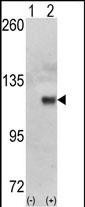
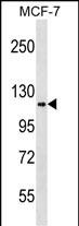
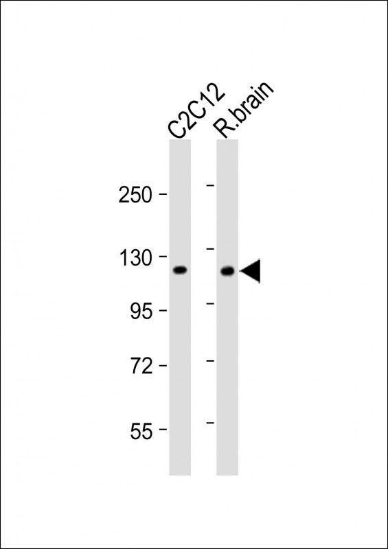
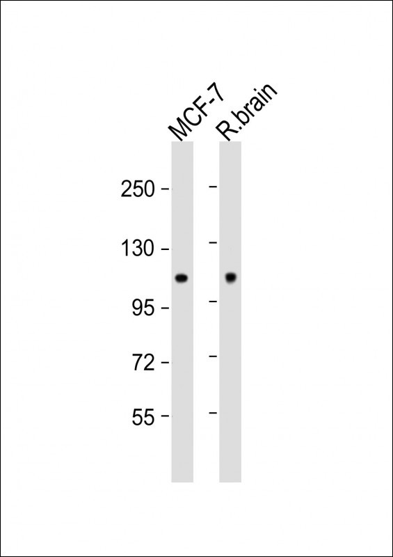
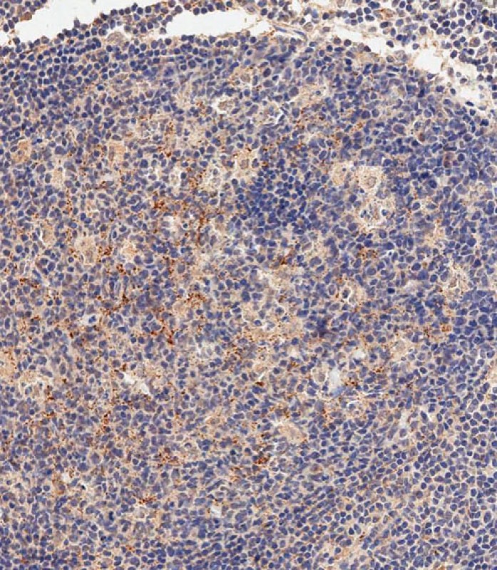
HK1 (Hexokinase) Antibody (N-term)
Purified Rabbit Polyclonal Antibody (Pab)
- SPECIFICATION
- CITATIONS: 2
- PROTOCOLS
- BACKGROUND

Application
| WB, IHC-P-Leica, E |
|---|---|
| Primary Accession | P19367 |
| Reactivity | Human, Rat |
| Host | Rabbit |
| Clonality | Polyclonal |
| Isotype | Rabbit IgG |
| Calculated MW | 102486 Da |
| Antigen Region | 78-108 aa |
| Gene ID | 3098 |
|---|---|
| Other Names | Hexokinase-1, Brain form hexokinase, Hexokinase type I, HK I, HK1 |
| Target/Specificity | This HK1 (Hexokinase) antibody is generated from rabbits immunized with a KLH conjugated synthetic peptide between 78-108 amino acids from the N-terminal region of human HK1 (Hexokinase). |
| Dilution | WB~~1:1000 IHC-P-Leica~~1:500 E~~Use at an assay dependent concentration. |
| Format | Purified polyclonal antibody supplied in PBS with 0.09% (W/V) sodium azide. This antibody is purified through a protein A column, followed by peptide affinity purification. |
| Storage | Maintain refrigerated at 2-8°C for up to 2 weeks. For long term storage store at -20°C in small aliquots to prevent freeze-thaw cycles. |
| Precautions | HK1 (Hexokinase) Antibody (N-term) is for research use only and not for use in diagnostic or therapeutic procedures. |
| Name | HK1 (HGNC:4922) |
|---|---|
| Function | Catalyzes the phosphorylation of various hexoses, such as D- glucose, D-glucosamine, D-fructose, D-mannose and 2-deoxy-D-glucose, to hexose 6-phosphate (D-glucose 6-phosphate, D-glucosamine 6-phosphate, D-fructose 6-phosphate, D-mannose 6-phosphate and 2-deoxy-D-glucose 6- phosphate, respectively) (PubMed:1637300, PubMed:25316723, PubMed:27374331). Does not phosphorylate N-acetyl-D-glucosamine (PubMed:27374331). Mediates the initial step of glycolysis by catalyzing phosphorylation of D-glucose to D-glucose 6-phosphate (By similarity). Involved in innate immunity and inflammation by acting as a pattern recognition receptor for bacterial peptidoglycan (PubMed:27374331). When released in the cytosol, N-acetyl-D-glucosamine component of bacterial peptidoglycan inhibits the hexokinase activity of HK1 and causes its dissociation from mitochondrial outer membrane, thereby activating the NLRP3 inflammasome (PubMed:27374331). |
| Cellular Location | Mitochondrion outer membrane; Peripheral membrane protein. Cytoplasm, cytosol. Note=The mitochondrial-binding peptide (MBP) region promotes association with the mitochondrial outer membrane (Probable). Dissociates from the mitochondrial outer membrane following inhibition by N-acetyl-D-glucosamine, leading to relocation to the cytosol (PubMed:27374331). |
| Tissue Location | Isoform 2: Erythrocyte specific (Ref.6). Isoform 3: Testis-specific (PubMed:10978502). Isoform 4: Testis-specific (PubMed:10978502). {ECO:0000269|PubMed:10978502, ECO:0000269|Ref.6} |

Provided below are standard protocols that you may find useful for product applications.
Background
Hexokinases phosphorylate glucose to produce glucose-6-phosphate, thus committing glucose to the glycolytic pathway. The hexokinase gene encodes a ubiquitous form of hexokinase which localizes to the outer membrane of mitochondria. Mutations in this gene have been associated with hemolytic anemia due to hexokinase deficiency. Alternative splicing of the hexokinase gene results in five transcript variants which encode different isoforms, some of which are tissue-specific. Each isoform has a distinct N-terminus; the remainder of the protein is identical among all the isoforms. HK1 encodes the ubiquitously expressed isoform. Its 5' end includes an exon which is unique to this transcript and which encodes a distinct N-terminus that contains the porin binding domain (PBD). The porin binding domain mediates association with the mitochondrial membrane.
References
van Wijk, R., et al., Blood 101(1):345-347 (2003).
Murakami, K., et al., Acta Haematol. 108(4):204-209 (2002).
Murakami, K., et al., Mol. Genet. Metab. 67(2):118-130 (1999).
Aleshin, A.E., et al., Structure 6(1):39-50 (1998).
Ruzzo, A., et al., Blood 91(1):363-364 (1998).
If you have used an Abcepta product and would like to share how it has performed, please click on the "Submit Review" button and provide the requested information. Our staff will examine and post your review and contact you if needed.
If you have any additional inquiries please email technical services at tech@abcepta.com.
Ordering Information
Other Products
Shipping Information
