


 Foundational characteristics of cancer include proliferation, angiogenesis, migration, evasion of apoptosis, and cellular immortality. Find key markers for these cellular processes and antibodies to detect them.
 The SUMOplot™ Analysis Program predicts and scores sumoylation sites in your protein. SUMOylation is a post-translational modification involved in various cellular processes, such as nuclear-cytosolic transport, transcriptional regulation, apoptosis, protein stability, response to stress, and progression through the cell cycle.
 The Autophagy Receptor Motif Plotter predicts and scores autophagy receptor binding sites in your protein. Identifying proteins connected to this pathway is critical to understanding the role of autophagy in physiological as well as pathological processes such as development, differentiation, neurodegenerative diseases, stress, infection, and cancer.
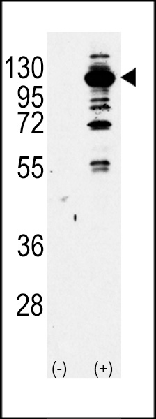
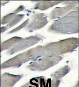
PI3KCG Antibody (Center)
Purified Rabbit Polyclonal Antibody (Pab)
- SPECIFICATION
- CITATIONS
- PROTOCOLS
- BACKGROUND

Application
| IHC-P, WB, E |
|---|---|
| Primary Accession | P48736 |
| Reactivity | Human |
| Host | Rabbit |
| Clonality | Polyclonal |
| Isotype | Rabbit IgG |
| Calculated MW | 126454 Da |
| Antigen Region | 518-547 aa |
| Gene ID | 5294 |
|---|---|
| Other Names | Phosphatidylinositol 4, 5-bisphosphate 3-kinase catalytic subunit gamma isoform, PI3-kinase subunit gamma, PI3K-gamma, PI3Kgamma, PtdIns-3-kinase subunit gamma, Phosphatidylinositol 4, 5-bisphosphate 3-kinase 110 kDa catalytic subunit gamma, PtdIns-3-kinase subunit p110-gamma, p110gamma, Phosphoinositide-3-kinase catalytic gamma polypeptide, Serine/threonine protein kinase PIK3CG, p120-PI3K, PIK3CG |
| Target/Specificity | This PI3KCG antibody is generated from rabbits immunized with a KLH conjugated synthetic peptide between 518-547 amino acids from the Central region of human PI3KCG. |
| Dilution | IHC-P~~1:10~50 WB~~1:1000 E~~Use at an assay dependent concentration. |
| Format | Purified polyclonal antibody supplied in PBS with 0.09% (W/V) sodium azide. This antibody is prepared by Saturated Ammonium Sulfate (SAS) precipitation followed by dialysis against PBS. |
| Storage | Maintain refrigerated at 2-8°C for up to 2 weeks. For long term storage store at -20°C in small aliquots to prevent freeze-thaw cycles. |
| Precautions | PI3KCG Antibody (Center) is for research use only and not for use in diagnostic or therapeutic procedures. |
| Name | PIK3CG |
|---|---|
| Function | Phosphoinositide-3-kinase (PI3K) that phosphorylates PtdIns(4,5)P2 (Phosphatidylinositol 4,5-bisphosphate) to generate phosphatidylinositol 3,4,5-trisphosphate (PIP3). PIP3 plays a key role by recruiting PH domain-containing proteins to the membrane, including AKT1 and PDPK1, activating signaling cascades involved in cell growth, survival, proliferation, motility and morphology. Links G-protein coupled receptor activation to PIP3 production. Involved in immune, inflammatory and allergic responses. Modulates leukocyte chemotaxis to inflammatory sites and in response to chemoattractant agents. May control leukocyte polarization and migration by regulating the spatial accumulation of PIP3 and by regulating the organization of F-actin formation and integrin-based adhesion at the leading edge. Controls motility of dendritic cells. Together with PIK3CD is involved in natural killer (NK) cell development and migration towards the sites of inflammation. Participates in T-lymphocyte migration. Regulates T- lymphocyte proliferation, activation, and cytokine production. Together with PIK3CD participates in T-lymphocyte development. Required for B- lymphocyte development and signaling. Together with PIK3CD participates in neutrophil respiratory burst. Together with PIK3CD is involved in neutrophil chemotaxis and extravasation. Together with PIK3CB promotes platelet aggregation and thrombosis. Regulates alpha-IIb/beta-3 integrins (ITGA2B/ ITGB3) adhesive function in platelets downstream of P2Y12 through a lipid kinase activity-independent mechanism. May have also a lipid kinase activity-dependent function in platelet aggregation. Involved in endothelial progenitor cell migration. Negative regulator of cardiac contractility. Modulates cardiac contractility by anchoring protein kinase A (PKA) and PDE3B activation, reducing cAMP levels. Regulates cardiac contractility also by promoting beta-adrenergic receptor internalization by binding to GRK2 and by non- muscle tropomyosin phosphorylation. Also has serine/threonine protein kinase activity: both lipid and protein kinase activities are required for beta-adrenergic receptor endocytosis. May also have a scaffolding role in modulating cardiac contractility. Contributes to cardiac hypertrophy under pathological stress. Through simultaneous binding of PDE3B to RAPGEF3 and PIK3R6 is assembled in a signaling complex in which the PI3K gamma complex is activated by RAPGEF3 and which is involved in angiogenesis. In neutrophils, participates in a phospholipase C-activating N-formyl peptide-activated GPCR (G protein- coupled receptor) signaling pathway downstream of RASGRP4-mediated Ras- activation, to promote neutrophil functional responses (By similarity). |
| Cellular Location | Cytoplasm. Cell membrane |
| Tissue Location | Pancreas, skeletal muscle, liver and heart. |

Thousands of laboratories across the world have published research that depended on the performance of antibodies from Abcepta to advance their research. Check out links to articles that cite our products in major peer-reviewed journals, organized by research category.
info@abcepta.com, and receive a free "I Love Antibodies" mug.

Provided below are standard protocols that you may find useful for product applications.
Background
PIK3CG belongs to the pi3/pi4-kinase family of proteins. It is an enzyme that phosphorylates phosphoinositides on the 3-hydroxyl group of the inositol ring. It is an important modulator of extracellular signals, including those elicited by E-cadherin-mediated cell-cell adhesion, which plays an important role in maintenance of the structural and functional integrity of epithelia. In addition to its role in promoting assembly of adherens junctions, the protein is thought to play a pivotal role in the regulation of cytotoxicity in NK cells. The gene for this protein is located in a commonly deleted segment of chromosome 7 previously identified in myeloid leukemias.
References
Andreozzi, F., et al., Endocrinology 145(6):2845-2857 (2004).
Reddy, S.A., et al., Biochem. Biophys. Res. Commun. 316(4):1022-1028 (2004).
Osaki, M., et al., J. Cancer Res. Clin. Oncol. 130(1):8-14 (2004).
Khan, N.A., et al., J. Neurovirol. 9(6):584-593 (2003).
Theberge, J.F., et al., Arch. Biochem. Biophys. 420(1):9-17 (2003).
If you have used an Abcepta product and would like to share how it has performed, please click on the "Submit Review" button and provide the requested information. Our staff will examine and post your review and contact you if needed.
If you have any additional inquiries please email technical services at tech@abcepta.com.
Ordering Information
Other Products
Shipping Information
