


 Foundational characteristics of cancer include proliferation, angiogenesis, migration, evasion of apoptosis, and cellular immortality. Find key markers for these cellular processes and antibodies to detect them.
 The SUMOplot™ Analysis Program predicts and scores sumoylation sites in your protein. SUMOylation is a post-translational modification involved in various cellular processes, such as nuclear-cytosolic transport, transcriptional regulation, apoptosis, protein stability, response to stress, and progression through the cell cycle.
 The Autophagy Receptor Motif Plotter predicts and scores autophagy receptor binding sites in your protein. Identifying proteins connected to this pathway is critical to understanding the role of autophagy in physiological as well as pathological processes such as development, differentiation, neurodegenerative diseases, stress, infection, and cancer.
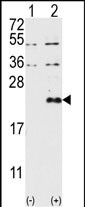
DUSP13-M1 Antibody (N-term)
Purified Rabbit Polyclonal Antibody (Pab)
- SPECIFICATION
- CITATIONS: 1
- PROTOCOLS
- BACKGROUND

Application
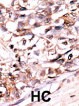
| IHC-P, WB, E |
|---|---|
| Primary Accession | Q9UII6 |
| Reactivity | Human |
| Host | Rabbit |
| Clonality | Polyclonal |
| Isotype | Rabbit IgG |
| Calculated MW | 22149 Da |
| Antigen Region | 1-30 aa |
| Gene ID | 51207 |
|---|---|
| Other Names | Dual specificity protein phosphatase 13 isoform B, DUSP13B, Dual specificity phosphatase SKRP4, Testis- and skeletal-muscle-specific DSP, DUSP13, DUSP13B, TMDP |
| Target/Specificity | This DUSP13-M1 antibody is generated from rabbits immunized with a KLH conjugated synthetic peptide between 1-30 amino acids from the N-terminal region of human DUSP13-M1. |
| Dilution | IHC-P~~1:50~100 WB~~1:1000 E~~Use at an assay dependent concentration. |
| Format | Purified polyclonal antibody supplied in PBS with 0.09% (W/V) sodium azide. This antibody is prepared by Saturated Ammonium Sulfate (SAS) precipitation followed by dialysis against PBS. |
| Storage | Maintain refrigerated at 2-8°C for up to 2 weeks. For long term storage store at -20°C in small aliquots to prevent freeze-thaw cycles. |
| Precautions | DUSP13-M1 Antibody (N-term) is for research use only and not for use in diagnostic or therapeutic procedures. |
| Name | DUSP13B (HGNC:19681) |
|---|---|
| Synonyms | DUSP13, SKRP4, TMDP |
| Function | Dual specificity phosphatase that dephosphorylates MAPK8/JNK and MAPK14/p38, but not MAPK1/ERK2, in vitro (PubMed:21360282). Exhibits intrinsic phosphatase activity towards both phospho- seryl/threonyl and -tyrosyl residues, with similar specific activities in vitro (PubMed:10585869). |
| Tissue Location | Highly expressed in the testis (at protein level) (PubMed:10585869, PubMed:15252030). Also found in the skeletal muscle (PubMed:15252030). |

Provided below are standard protocols that you may find useful for product applications.
Background
Dual-specificity phosphatases, a subfamily of protein-tyrosine phosphatases, play important roles in signal transduction, cell cycle progression, and tumor suppression. The cDNA encoding a novel phosphatase, PIR1, phosphatase that interacts with RNA/RNP complex 1. Sequence analysis revealed that the predicted 329-amino acid protein has homology to several dual-specificity phosphatases and contains 2 stretches of arginine-rich sequence similar to those found in some RNA-binding proteins. In vitro, recombinant protein displays protein-tyrosine phosphatase activity and binds directly to RNA.
References
Nakamura K., Shima H., Watanabe M., Haneji T., Kikuchi K.Biochem. J. 344:819-825(1999).
Deloukas et al. Nature 429:375-381(2004).
Strausberg et al. Proc. Natl. Acad. Sci. U.S.A. 99:16899-16903(2002).
If you have used an Abcepta product and would like to share how it has performed, please click on the "Submit Review" button and provide the requested information. Our staff will examine and post your review and contact you if needed.
If you have any additional inquiries please email technical services at tech@abcepta.com.
Ordering Information
Shipping Information
