


 Foundational characteristics of cancer include proliferation, angiogenesis, migration, evasion of apoptosis, and cellular immortality. Find key markers for these cellular processes and antibodies to detect them.
 The SUMOplot™ Analysis Program predicts and scores sumoylation sites in your protein. SUMOylation is a post-translational modification involved in various cellular processes, such as nuclear-cytosolic transport, transcriptional regulation, apoptosis, protein stability, response to stress, and progression through the cell cycle.
 The Autophagy Receptor Motif Plotter predicts and scores autophagy receptor binding sites in your protein. Identifying proteins connected to this pathway is critical to understanding the role of autophagy in physiological as well as pathological processes such as development, differentiation, neurodegenerative diseases, stress, infection, and cancer.
> home > Products > Primary Antibodies > Antibody Collections > Inflammatory response > CCL3 Antibody (C-term)
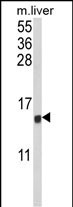
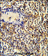
CCL3 Antibody (C-term)
Affinity Purified Rabbit Polyclonal Antibody (Pab)
- SPECIFICATION
- CITATIONS: 2
- PROTOCOLS
- BACKGROUND

Application
| IHC-P, WB, E |
|---|---|
| Primary Accession | P10147 |
| Other Accession | P16619 |
| Reactivity | Human, Mouse |
| Host | Rabbit |
| Clonality | Polyclonal |
| Isotype | Rabbit IgG |
| Calculated MW | 10085 Da |
| Antigen Region | 59-87 aa |
| Gene ID | 6348 |
|---|---|
| Other Names | C-C motif chemokine 3, G0/G1 switch regulatory protein 19-1, Macrophage inflammatory protein 1-alpha, MIP-1-alpha, PAT 4641, SIS-beta, Small-inducible cytokine A3, Tonsillar lymphocyte LD78 alpha protein, MIP-1-alpha(4-69), LD78-alpha(4-69), CCL3, G0S19-1, MIP1A, SCYA3 |
| Target/Specificity | This CCL3 antibody is generated from rabbits immunized with a KLH conjugated synthetic peptide between 59-87 amino acids from the C-terminal region of human CCL3. |
| Dilution | IHC-P~~1:50~100 WB~~1:1000 E~~Use at an assay dependent concentration. |
| Format | Purified polyclonal antibody supplied in PBS with 0.09% (W/V) sodium azide. This antibody is purified through a protein A column, followed by peptide affinity purification. |
| Storage | Maintain refrigerated at 2-8°C for up to 2 weeks. For long term storage store at -20°C in small aliquots to prevent freeze-thaw cycles. |
| Precautions | CCL3 Antibody (C-term) is for research use only and not for use in diagnostic or therapeutic procedures. |
| Name | CCL3 |
|---|---|
| Synonyms | G0S19-1, MIP1A, SCYA3 |
| Function | Monokine with inflammatory and chemokinetic properties. Binds to CCR1, CCR4 and CCR5. One of the major HIV-suppressive factors produced by CD8+ T-cells. Recombinant MIP-1-alpha induces a dose- dependent inhibition of different strains of HIV-1, HIV-2, and simian immunodeficiency virus (SIV). |
| Cellular Location | Secreted. |
Research Areas
Citations ( 0 )

Application Protocols
Provided below are standard protocols that you may find useful for product applications.
Background
Macrophage inflammatory protein-1 is a so-called monokine that is involved in the acute inflammatory state in the recruitment and activation of polymorphonuclear leukocytes.
References
Hirashima,M., et.al., DNA Seq. 3 (4), 203-212 (1992) Blum,S., et.al., DNA Cell Biol. 9 (8), 589-602 (1990)
Abcepta welcomes feedback from its customers.
If you have used an Abcepta product and would like to share how it has performed, please click on the "Submit Review" button and provide the requested information. Our staff will examine and post your review and contact you if needed.
If you have any additional inquiries please email technical services at tech@abcepta.com.
$ 75.00

⚠️ Limit: 5 vials per order for discount.
$ 192.50
⚠️ Limit: 5 vials per order for discount.
Cat# AP8527b
Ordering Information
United States
AlbaniaAustraliaAustriaBelgiumBosnia & HerzegovinaBrazilBulgariaCanadaCentral AmericaChinaCroatiaCyprusCzech RepublicDenmarkEstoniaFinlandFranceGermanyGreeceHong KongHungaryIcelandIndiaIndonesiaIrelandIsraelItalyJapanLatviaLithuaniaLuxembourgMacedoniaMalaysiaMaltaMexicoNetherlandsNew ZealandNorwayPakistanPolandPortugalRomaniaSerbiaSingaporeSlovakiaSloveniaSouth AfricaSouth KoreaSpainSwedenSwitzerlandTaiwanTurkeyUnited KingdomUnited StatesVietnamWorldwideOthers
USA Headquarters
(888) 735-7227 / (858) 622-0099 or (858) 875-1900
Other Products
Shipping Information
Domestic orders (in stock items)
Shipped out the same day. Orders placed after 1 PM (PST) will ship out the next business day.
International orders
Contact your local distributors
