


 Foundational characteristics of cancer include proliferation, angiogenesis, migration, evasion of apoptosis, and cellular immortality. Find key markers for these cellular processes and antibodies to detect them.
 The SUMOplot™ Analysis Program predicts and scores sumoylation sites in your protein. SUMOylation is a post-translational modification involved in various cellular processes, such as nuclear-cytosolic transport, transcriptional regulation, apoptosis, protein stability, response to stress, and progression through the cell cycle.
 The Autophagy Receptor Motif Plotter predicts and scores autophagy receptor binding sites in your protein. Identifying proteins connected to this pathway is critical to understanding the role of autophagy in physiological as well as pathological processes such as development, differentiation, neurodegenerative diseases, stress, infection, and cancer.
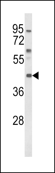
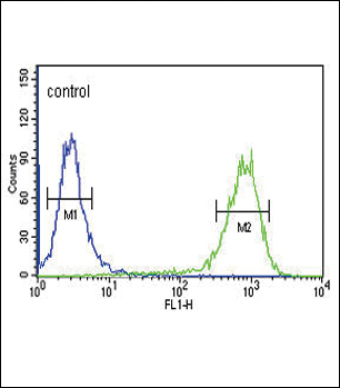
MC3R Antibody (Center)
Affinity Purified Rabbit Polyclonal Antibody (Pab)
- SPECIFICATION
- CITATIONS
- PROTOCOLS
- BACKGROUND

Application
| FC, WB, E |
|---|---|
| Primary Accession | P41968 |
| Reactivity | Human |
| Host | Rabbit |
| Clonality | Polyclonal |
| Isotype | Rabbit IgG |
| Calculated MW | 36043 Da |
| Antigen Region | 86-112 aa |
| Gene ID | 4159 |
|---|---|
| Other Names | Melanocortin receptor 3, MC3-R, MC3R |
| Target/Specificity | This MC3R antibody is generated from rabbits immunized with a KLH conjugated synthetic peptide between 86-112 amino acids from the Central region of human MC3R. |
| Dilution | FC~~1:10~50 WB~~1:1000 E~~Use at an assay dependent concentration. |
| Format | Purified polyclonal antibody supplied in PBS with 0.09% (W/V) sodium azide. This antibody is purified through a protein A column, followed by peptide affinity purification. |
| Storage | Maintain refrigerated at 2-8°C for up to 2 weeks. For long term storage store at -20°C in small aliquots to prevent freeze-thaw cycles. |
| Precautions | MC3R Antibody (Center) is for research use only and not for use in diagnostic or therapeutic procedures. |
| Name | MC3R (HGNC:6931) |
|---|---|
| Function | G protein-coupled receptor for melanocyte-stimulating hormones (alpha, beta, and gamma-MSH) and corticotropin/ACTH, which are peptide products of the POMC precursor (PubMed:37524700, PubMed:8463333). Upon activation, couples to G(s) protein, stimulating adenylate cyclase and the cAMP-dependent signaling pathway, which contributes to the regulation of energy homeostasis (PubMed:18231126, PubMed:37524700, PubMed:8463333). Required for expression of anticipatory patterns of activity and wakefulness during periods of limited nutrient availability and for the normal regulation of circadian clock activity in the brain (By similarity). Binding of the Agouti-related protein/AGPR antagonist precludes alpha-MSH-induced signaling, blocking cAMP production (PubMed:9311920). |
| Cellular Location | Cell membrane; Multi-pass membrane protein. |
| Tissue Location | Brain, placental, and gut tissues. |

Thousands of laboratories across the world have published research that depended on the performance of antibodies from Abcepta to advance their research. Check out links to articles that cite our products in major peer-reviewed journals, organized by research category.
info@abcepta.com, and receive a free "I Love Antibodies" mug.

Provided below are standard protocols that you may find useful for product applications.
Background
MC3R is a G-protein-coupled receptor for melanocyte-stimulating hormone and adrenocorticotropic hormone that is expressed in tissues other than the adrenal cortex and melanocytes. This gene maps to the same region as the locus for benign neonatal epilepsy.
References
Magenis,R.E.,et.al., Mamm. Genome 5 (8), 503-508 (1994)
Konda,Y., et.al., J. Biol. Chem. 269 (18), 13162-13166 (1994)
If you have used an Abcepta product and would like to share how it has performed, please click on the "Submit Review" button and provide the requested information. Our staff will examine and post your review and contact you if needed.
If you have any additional inquiries please email technical services at tech@abcepta.com.
Ordering Information
Other Products
Shipping Information
