


 Foundational characteristics of cancer include proliferation, angiogenesis, migration, evasion of apoptosis, and cellular immortality. Find key markers for these cellular processes and antibodies to detect them.
 The SUMOplot™ Analysis Program predicts and scores sumoylation sites in your protein. SUMOylation is a post-translational modification involved in various cellular processes, such as nuclear-cytosolic transport, transcriptional regulation, apoptosis, protein stability, response to stress, and progression through the cell cycle.
 The Autophagy Receptor Motif Plotter predicts and scores autophagy receptor binding sites in your protein. Identifying proteins connected to this pathway is critical to understanding the role of autophagy in physiological as well as pathological processes such as development, differentiation, neurodegenerative diseases, stress, infection, and cancer.
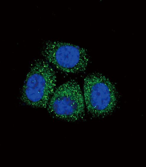
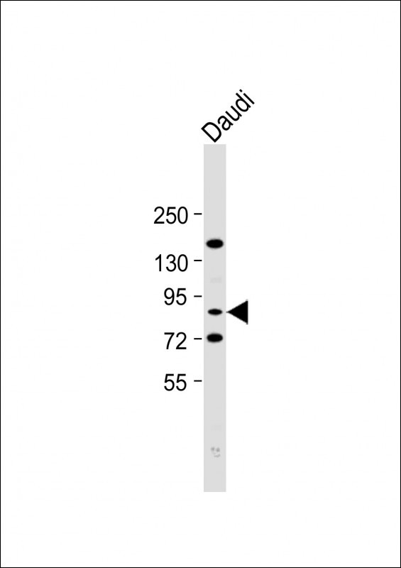
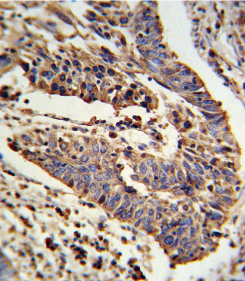
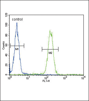
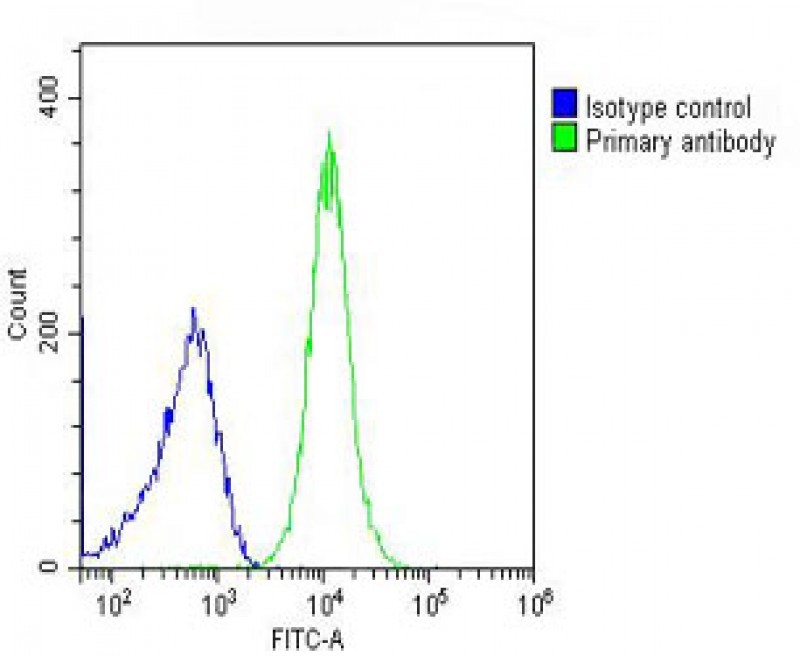
SCNN1A Antibody (Center)
Affinity Purified Rabbit Polyclonal Antibody (Pab)
- SPECIFICATION
- CITATIONS
- PROTOCOLS
- BACKGROUND

Application
| FC, IF, IHC-P, WB, E |
|---|---|
| Primary Accession | P37088 |
| Reactivity | Human |
| Host | Rabbit |
| Clonality | Polyclonal |
| Isotype | Rabbit IgG |
| Calculated MW | 75704 Da |
| Antigen Region | 365-391 aa |
| Gene ID | 6337 |
|---|---|
| Other Names | Amiloride-sensitive sodium channel subunit alpha, Alpha-NaCH, Epithelial Na(+) channel subunit alpha, Alpha-ENaC, ENaCA, Nonvoltage-gated sodium channel 1 subunit alpha, SCNEA, SCNN1A, SCNN1 |
| Target/Specificity | This SCNN1A antibody is generated from rabbits immunized with a KLH conjugated synthetic peptide between 365-391 amino acids from the Central region of human SCNN1A. |
| Dilution | FC~~1:25 IF~~1:10~50 IHC-P~~1:10~50 WB~~1:2000 E~~Use at an assay dependent concentration. |
| Format | Purified polyclonal antibody supplied in PBS with 0.09% (W/V) sodium azide. This antibody is purified through a protein A column, followed by peptide affinity purification. |
| Storage | Maintain refrigerated at 2-8°C for up to 2 weeks. For long term storage store at -20°C in small aliquots to prevent freeze-thaw cycles. |
| Precautions | SCNN1A Antibody (Center) is for research use only and not for use in diagnostic or therapeutic procedures. |
| Name | SCNN1A (HGNC:10599) |
|---|---|
| Function | This is one of the three pore-forming subunits of the heterotrimeric epithelial sodium channel (ENaC), a critical regulator of sodium balance and fluid homeostasis (PubMed:30251954, PubMed:32729833, PubMed:8023962, PubMed:8278374, PubMed:9792722). ENaC operates in epithelial tissues, where it mediates the electrodiffusion of sodium ions from extracellular fluid through the apical membrane of cells, with water following osmotically (PubMed:24124190, PubMed:28710092, PubMed:8278374). It plays a key role in maintaining sodium homeostasis through electrogenic sodium reabsorption in the kidneys (PubMed:12107247). Additionally, ENaC is essential for airway surface liquid homeostasis, which is crucial for proper mucus clearance (PubMed:24124190, PubMed:28710092). |
| Cellular Location | Apical cell membrane; Multi-pass membrane protein. Cell projection, cilium. Cytoplasmic granule. Cytoplasm Cytoplasmic vesicle, secretory vesicle, acrosome {ECO:0000250|UniProtKB:P37089}. Cell projection, cilium, flagellum {ECO:0000250|UniProtKB:P37089}. Note=In the oviduct and bronchus, located on cilia in multi-ciliated cells. In endometrial non-ciliated epithelial cells, restricted to apical surfaces. In epidermis, located nearly uniformly in the cytoplasm in a granular distribution (PubMed:28130590). In sebaceous glands, observed only in the cytoplasmic space in between the lipid vesicles (PubMed:28130590). In eccrine sweat glands, mainly located at the apical surface of the cells facing the lumen (PubMed:28130590). In skin, in arrector pili muscle cells and in adipocytes, located in the cytoplasm and colocalized with actin fibers (PubMed:28130590). In spermatogonia, spermatocytes and round spermatids, located in the cytoplasm (By similarity). Prior to spermiation, location shifts from the cytoplasm to the spermatid tail (By similarity). In spermatozoa, localizes at the acrosome and the central region of the sperm flagellum (By similarity) {ECO:0000250|UniProtKB:P37089, ECO:0000269|PubMed:22207244, ECO:0000269|PubMed:24124190, ECO:0000269|PubMed:28130590} |
| Tissue Location | Expressed in the female reproductive tract, from the fimbrial end of the fallopian tube to the endometrium (at protein level) (PubMed:22207244). Expressed in kidney (at protein level). In the respiratory tract, expressed in the bronchial epithelium (at protein level). Highly expressed in lung. Detected at intermediate levels in pancreas and liver, and at low levels in heart and placenta (PubMed:22207244). in skin, expressed in keratinocytes, melanocytes and Merkel cells of the epidermal sub-layers, stratum basale, stratum spinosum and stratum granulosum (at protein level) (PubMed:28130590) Expressed in the outer root sheath of the hair follicles (at protein level) (PubMed:28130590). Detected in both peripheral and central cells of the sebaceous gland (at protein level) (PubMed:28130590). Expressed by eccrine sweat glands (at protein level) (PubMed:28130590). In skin, also expressed by arrector pili muscle cells and intradermal adipocytes (PubMed:28130590). Isoform 1 and isoform 2 predominate in all tissues |

Thousands of laboratories across the world have published research that depended on the performance of antibodies from Abcepta to advance their research. Check out links to articles that cite our products in major peer-reviewed journals, organized by research category.
info@abcepta.com, and receive a free "I Love Antibodies" mug.

Provided below are standard protocols that you may find useful for product applications.
Background
Nonvoltage-gated, amiloride-sensitive, sodium channels control fluid and electrolyte transport across epithelia in many organs. These channels are heteromeric complexes consisting of 3 subunits: alpha, beta, and gamma. This gene encodes the alpha subunit, and mutations in this gene have been sociated with pseudohypoaldosteronism type 1 (PHA1), a rare salt wasting disease resulting from target organ unresponsiveness to mineralocorticoids.
References
Azad A.K., et.al., Hum. Mutat. 30:1093-1103(2009).
If you have used an Abcepta product and would like to share how it has performed, please click on the "Submit Review" button and provide the requested information. Our staff will examine and post your review and contact you if needed.
If you have any additional inquiries please email technical services at tech@abcepta.com.
Ordering Information
Other Products
Shipping Information
