


 Foundational characteristics of cancer include proliferation, angiogenesis, migration, evasion of apoptosis, and cellular immortality. Find key markers for these cellular processes and antibodies to detect them.
 The SUMOplot™ Analysis Program predicts and scores sumoylation sites in your protein. SUMOylation is a post-translational modification involved in various cellular processes, such as nuclear-cytosolic transport, transcriptional regulation, apoptosis, protein stability, response to stress, and progression through the cell cycle.
 The Autophagy Receptor Motif Plotter predicts and scores autophagy receptor binding sites in your protein. Identifying proteins connected to this pathway is critical to understanding the role of autophagy in physiological as well as pathological processes such as development, differentiation, neurodegenerative diseases, stress, infection, and cancer.
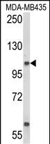
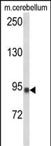
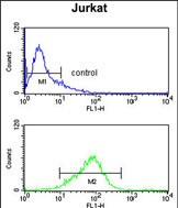
CASD1 Antibody (N-term)
Affinity Purified Rabbit Polyclonal Antibody (Pab)
- SPECIFICATION
- CITATIONS
- PROTOCOLS
- BACKGROUND

Application
| FC, WB, E |
|---|---|
| Primary Accession | Q96PB1 |
| Other Accession | Q7TN73 |
| Reactivity | Human, Mouse |
| Host | Rabbit |
| Clonality | Polyclonal |
| Isotype | Rabbit IgG |
| Calculated MW | 91680 Da |
| Antigen Region | 97-126 aa |
| Gene ID | 64921 |
|---|---|
| Other Names | CAS1 domain-containing protein 1, CASD1, C7orf12 |
| Target/Specificity | This CASD1 antibody is generated from rabbits immunized with a KLH conjugated synthetic peptide between 97-126 amino acids from the N-terminal region of human CASD1. |
| Dilution | FC~~1:10~50 WB~~1:1000 E~~Use at an assay dependent concentration. |
| Format | Purified polyclonal antibody supplied in PBS with 0.09% (W/V) sodium azide. This antibody is purified through a protein A column, followed by peptide affinity purification. |
| Storage | Maintain refrigerated at 2-8°C for up to 2 weeks. For long term storage store at -20°C in small aliquots to prevent freeze-thaw cycles. |
| Precautions | CASD1 Antibody (N-term) is for research use only and not for use in diagnostic or therapeutic procedures. |
| Name | CASD1 (HGNC:16014) |
|---|---|
| Synonyms | C7orf12 |
| Function | Key enzyme in the biosynthesis of O-acetylated (O-Ac) sialoglycans such as gangliosides O-AcGD3 and O-AcGD2, which affect various processes such as cell-cell interactions, host-pathogen recognition (PubMed:20947662, PubMed:26169044, PubMed:34208013). Catalyzes the transfer of an acetyl group from a donor, the acetyl- coenzyme-A molecule (acetyl-CoA), to the C7/8/9 OH-position of a sialic acid residue (PubMed:20947662, PubMed:26169044). The primary site of O- acetyl group transfer on sialic acid seems to depend on cell type and can be C7, from which the O-acetyl group could subsequently migrate to the C8 and then to the C9 position, or at C9 with possibility of migrating to the C8 and then to the C7 position (PubMed:20947662, PubMed:26169044). Together with ST8SIA1 (GD3 synthase) it increases the levels of ganglioside Ac-O-7-GD3 (PubMed:20947662). Can transfer the acetyl group from acetyl-CoA to free sialate (N-acetylneuraminate, Neu5Ac) in vitro, but has preferred substrate specificity for CMP- activated sialate (CMP-Neu5Ac), resulting in the formation of 9-O- acetylated CMP-Neu5Ac (CMP-Neu5,9Ac2) (PubMed:26169044). CMP-Neu5,9Ac2 may be used by sialyltransferases as a sialate donor for glycoconjugate acceptors such as ganglioside GD3 (PubMed:26169044). O-acetylation at position C9 of ganglioside GD3 can counteract the pro-apoptotic effects of the ganglioside GD3 in tumor cells (PubMed:12486096). |
| Cellular Location | Golgi apparatus membrane; Multi-pass membrane protein |
| Tissue Location | Highly expressed in peripheral B lymphocytes. |
Research Areas
Citations (0)

Thousands of laboratories across the world have published research that depended on the performance of antibodies from Abcepta to advance their research. Check out links to articles that cite our products in major peer-reviewed journals, organized by research category.
Submit your citation using an Abcepta antibody to
info@abcepta.com, and receive a free "I Love Antibodies" mug.
info@abcepta.com, and receive a free "I Love Antibodies" mug.

Application Protocols
Provided below are standard protocols that you may find useful for product applications.
References
Janbon,G.,et.al., Mol. Microbiol. 42 (2), 453-467 (2001)
Abcepta welcomes feedback from its customers.
If you have used an Abcepta product and would like to share how it has performed, please click on the "Submit Review" button and provide the requested information. Our staff will examine and post your review and contact you if needed.
If you have any additional inquiries please email technical services at tech@abcepta.com.
$ 150.00
$ 385.00
Cat# AP8857a
Ordering Information
United States
AlbaniaAustraliaAustriaBelgiumBosnia & HerzegovinaBrazilBulgariaCanadaCentral AmericaChinaCroatiaCyprusCzech RepublicDenmarkEstoniaFinlandFranceGermanyGreeceHong KongHungaryIcelandIndiaIndonesiaIrelandIsraelItalyJapanLatviaLithuaniaLuxembourgMacedoniaMalaysiaMaltaMexicoNetherlandsNew ZealandNorwayPakistanPolandPortugalRomaniaSerbiaSingaporeSlovakiaSloveniaSouth AfricaSouth KoreaSpainSwedenSwitzerlandTaiwanTurkeyUnited KingdomUnited StatesVietnamWorldwideOthers
USA Headquarters
(888) 735-7227 / (858) 622-0099 or (858) 875-1900
Shipping Information
Domestic orders (in stock items)
Shipped out the same day. Orders placed after 1 PM (PST) will ship out the next business day.
International orders
Contact your local distributors
