


 Foundational characteristics of cancer include proliferation, angiogenesis, migration, evasion of apoptosis, and cellular immortality. Find key markers for these cellular processes and antibodies to detect them.
 The SUMOplot™ Analysis Program predicts and scores sumoylation sites in your protein. SUMOylation is a post-translational modification involved in various cellular processes, such as nuclear-cytosolic transport, transcriptional regulation, apoptosis, protein stability, response to stress, and progression through the cell cycle.
 The Autophagy Receptor Motif Plotter predicts and scores autophagy receptor binding sites in your protein. Identifying proteins connected to this pathway is critical to understanding the role of autophagy in physiological as well as pathological processes such as development, differentiation, neurodegenerative diseases, stress, infection, and cancer.
DTX1 Antibody (Center)
Affinity Purified Rabbit Polyclonal Antibody (Pab)
- SPECIFICATION
- CITATIONS
- PROTOCOLS
- BACKGROUND

Application
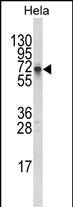
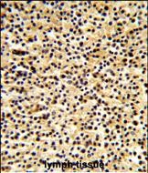
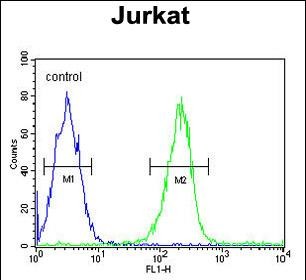
| FC, IHC-P, WB, E |
|---|---|
| Primary Accession | Q86Y01 |
| Other Accession | Q61010 |
| Reactivity | Human |
| Predicted | Mouse |
| Host | Rabbit |
| Clonality | Polyclonal |
| Isotype | Rabbit IgG |
| Calculated MW | 67368 Da |
| Antigen Region | 382-410 aa |
| Gene ID | 1840 |
|---|---|
| Other Names | E3 ubiquitin-protein ligase DTX1, 632-, Protein deltex-1, Deltex1, hDTX1, DTX1 |
| Target/Specificity | This DTX1 antibody is generated from rabbits immunized with a KLH conjugated synthetic peptide between 382-410 amino acids from the Central region of human DTX1. |
| Dilution | FC~~1:10~50 IHC-P~~1:50~100 WB~~1:1000 E~~Use at an assay dependent concentration. |
| Format | Purified polyclonal antibody supplied in PBS with 0.09% (W/V) sodium azide. This antibody is purified through a protein A column, followed by peptide affinity purification. |
| Storage | Maintain refrigerated at 2-8°C for up to 2 weeks. For long term storage store at -20°C in small aliquots to prevent freeze-thaw cycles. |
| Precautions | DTX1 Antibody (Center) is for research use only and not for use in diagnostic or therapeutic procedures. |
| Name | DTX1 |
|---|---|
| Function | Functions as a ubiquitin ligase protein in vivo, mediating ubiquitination and promoting degradation of MEKK1, suggesting that it may regulate the Notch pathway via some ubiquitin ligase activity (By similarity). Regulator of Notch signaling, a signaling pathway involved in cell-cell communications that regulates a broad spectrum of cell- fate determinations. Mainly acts as a positive regulator of Notch, but it also acts as a negative regulator, depending on the developmental and cell context. Mediates the antineural activity of Notch, possibly by inhibiting the transcriptional activation mediated by MATCH1. Involved in neurogenesis, lymphogenesis and myogenesis, and may also be involved in MZB (Marginal zone B) cell differentiation. Promotes B-cell development at the expense of T-cell development, suggesting that it can antagonize NOTCH1. |
| Cellular Location | Cytoplasm. Nucleus. Note=Predominantly cytoplasmic. Associates with endocytic vesicles. Partially nuclear |
| Tissue Location | Widely expressed. Strongly expressed in blood vessel. Also expressed in embryonic nervous system, pancreas, lung, adrenal gland, digestive tube and muscles. Expressed in MZB cells and developing B- and T-cells. |

Thousands of laboratories across the world have published research that depended on the performance of antibodies from Abcepta to advance their research. Check out links to articles that cite our products in major peer-reviewed journals, organized by research category.
info@abcepta.com, and receive a free "I Love Antibodies" mug.

Provided below are standard protocols that you may find useful for product applications.
Background
DTX1 was identified as encoding a positive regulator of the Notch-signaling pathway in Drosophila. The human gene encodes a protein of unknown function; however, it may play a role in basic helix-loop-helix transcription factor activity.
References
Wu,C.,et.al., Proteomics 7 (11), 1775-1785 (2007)
If you have used an Abcepta product and would like to share how it has performed, please click on the "Submit Review" button and provide the requested information. Our staff will examine and post your review and contact you if needed.
If you have any additional inquiries please email technical services at tech@abcepta.com.
Ordering Information
Other Products
Shipping Information
