


 Foundational characteristics of cancer include proliferation, angiogenesis, migration, evasion of apoptosis, and cellular immortality. Find key markers for these cellular processes and antibodies to detect them.
 The SUMOplot™ Analysis Program predicts and scores sumoylation sites in your protein. SUMOylation is a post-translational modification involved in various cellular processes, such as nuclear-cytosolic transport, transcriptional regulation, apoptosis, protein stability, response to stress, and progression through the cell cycle.
 The Autophagy Receptor Motif Plotter predicts and scores autophagy receptor binding sites in your protein. Identifying proteins connected to this pathway is critical to understanding the role of autophagy in physiological as well as pathological processes such as development, differentiation, neurodegenerative diseases, stress, infection, and cancer.
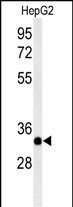
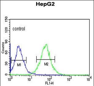
ACER3 Antibody (C-term)
Affinity Purified Rabbit Polyclonal Antibody (Pab)
- SPECIFICATION
- CITATIONS
- PROTOCOLS
- BACKGROUND

Application
| FC, WB, E |
|---|---|
| Primary Accession | Q9NUN7 |
| Other Accession | Q9D099 |
| Reactivity | Human, Mouse |
| Host | Rabbit |
| Clonality | Polyclonal |
| Isotype | Rabbit IgG |
| Calculated MW | 31552 Da |
| Antigen Region | 224-250 aa |
| Gene ID | 55331 |
|---|---|
| Other Names | Alkaline ceramidase 3, AlkCDase 3, Alkaline CDase 3, 351-, Alkaline dihydroceramidase SB89, Alkaline phytoceramidase, aPHC, ACER3, APHC, PHCA |
| Target/Specificity | This ACER3 antibody is generated from rabbits immunized with a KLH conjugated synthetic peptide between 224-250 amino acids from the C-terminal region of human ACER3. |
| Dilution | FC~~1:10~50 WB~~1:1000 E~~Use at an assay dependent concentration. |
| Format | Purified polyclonal antibody supplied in PBS with 0.09% (W/V) sodium azide. This antibody is purified through a protein A column, followed by peptide affinity purification. |
| Storage | Maintain refrigerated at 2-8°C for up to 2 weeks. For long term storage store at -20°C in small aliquots to prevent freeze-thaw cycles. |
| Precautions | ACER3 Antibody (C-term) is for research use only and not for use in diagnostic or therapeutic procedures. |
| Name | ACER3 |
|---|---|
| Synonyms | APHC, PHCA |
| Function | Endoplasmic reticulum and Golgi ceramidase that catalyzes the hydrolysis of unsaturated long-chain C18:1-, C20:1- and C20:4- ceramides, dihydroceramides and phytoceramides into sphingoid bases like sphingosine and free fatty acids at alkaline pH (PubMed:11356846, PubMed:20068046, PubMed:20207939, PubMed:26792856, PubMed:30575723). Ceramides, sphingosine, and its phosphorylated form sphingosine-1- phosphate are bioactive lipids that mediate cellular signaling pathways regulating several biological processes including cell proliferation, apoptosis and differentiation (PubMed:20068046). Controls the generation of sphingosine in erythrocytes, and thereby sphingosine-1- phosphate in plasma (PubMed:20207939). Through the regulation of ceramides and sphingosine-1-phosphate homeostasis in the brain may play a role in neurons survival and function (By similarity). By regulating the levels of pro-inflammatory ceramides in immune cells and tissues, may modulate the inflammatory response (By similarity). |
| Cellular Location | Endoplasmic reticulum membrane; Multi-pass membrane protein. Golgi apparatus membrane; Multi-pass membrane protein |
| Tissue Location | Ubiquitously expressed. Highly expressed in placenta (PubMed:11356846). Expressed in erythrocytes (PubMed:20207939). |

Thousands of laboratories across the world have published research that depended on the performance of antibodies from Abcepta to advance their research. Check out links to articles that cite our products in major peer-reviewed journals, organized by research category.
info@abcepta.com, and receive a free "I Love Antibodies" mug.

Provided below are standard protocols that you may find useful for product applications.
Background
ACER3 hydrolyzes only phytoceramide into phytosphingosine and free fatty acid. Does not have reverse activity.
References
Wheeler,H.E., et.al., PLoS Genet. 5 (10), E1000685 (2009)
Mao,C. et.al., Biochim. Biophys. Acta 1781 (9), 424-434 (2008)
If you have used an Abcepta product and would like to share how it has performed, please click on the "Submit Review" button and provide the requested information. Our staff will examine and post your review and contact you if needed.
If you have any additional inquiries please email technical services at tech@abcepta.com.
Ordering Information
Other Products
Shipping Information
