


 Foundational characteristics of cancer include proliferation, angiogenesis, migration, evasion of apoptosis, and cellular immortality. Find key markers for these cellular processes and antibodies to detect them.
 The SUMOplot™ Analysis Program predicts and scores sumoylation sites in your protein. SUMOylation is a post-translational modification involved in various cellular processes, such as nuclear-cytosolic transport, transcriptional regulation, apoptosis, protein stability, response to stress, and progression through the cell cycle.
 The Autophagy Receptor Motif Plotter predicts and scores autophagy receptor binding sites in your protein. Identifying proteins connected to this pathway is critical to understanding the role of autophagy in physiological as well as pathological processes such as development, differentiation, neurodegenerative diseases, stress, infection, and cancer.
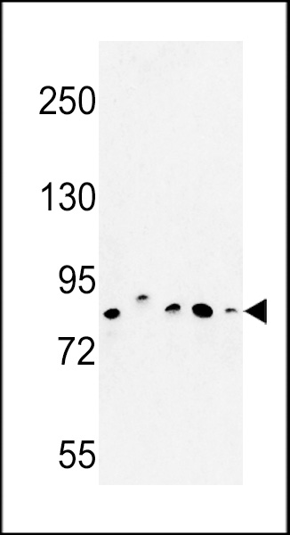
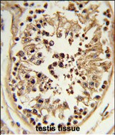
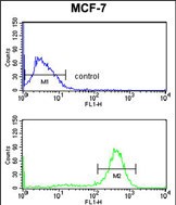
CHPF Antibody (Center)
Affinity Purified Rabbit Polyclonal Antibody (Pab)
- SPECIFICATION
- CITATIONS
- PROTOCOLS
- BACKGROUND

Application
| FC, IHC-P, WB, E |
|---|---|
| Primary Accession | Q8IZ52 |
| Reactivity | Human, Mouse |
| Host | Rabbit |
| Clonality | Polyclonal |
| Isotype | Rabbit IgG |
| Calculated MW | 85467 Da |
| Antigen Region | 327-354 aa |
| Gene ID | 79586 |
|---|---|
| Other Names | Chondroitin sulfate synthase 2, Chondroitin glucuronyltransferase 2, Chondroitin-polymerizing factor, ChPF, Glucuronosyl-N-acetylgalactosaminyl-proteoglycan 4-beta-N-acetylgalactosaminyltransferase II, N-acetylgalactosaminyl-proteoglycan 3-beta-glucuronosyltransferase II, N-acetylgalactosaminyltransferase 2, CHPF, CSS2 |
| Target/Specificity | This CHPF antibody is generated from rabbits immunized with a KLH conjugated synthetic peptide between 327-354 amino acids from the Central region of human CHPF. |
| Dilution | FC~~1:10~50 IHC-P~~1:50~100 WB~~1:1000 E~~Use at an assay dependent concentration. |
| Format | Purified polyclonal antibody supplied in PBS with 0.09% (W/V) sodium azide. This antibody is purified through a protein A column, followed by peptide affinity purification. |
| Storage | Maintain refrigerated at 2-8°C for up to 2 weeks. For long term storage store at -20°C in small aliquots to prevent freeze-thaw cycles. |
| Precautions | CHPF Antibody (Center) is for research use only and not for use in diagnostic or therapeutic procedures. |
| Name | CHPF (HGNC:24291) |
|---|---|
| Synonyms | CSS2 |
| Function | Has both beta-1,3-glucuronic acid and beta-1,4-N- acetylgalactosamine transferase activity. Transfers glucuronic acid (GlcUA) from UDP-GlcUA and N-acetylgalactosamine (GalNAc) from UDP- GalNAc to the non-reducing end of the elongating chondroitin polymer. Seems to act as a specific activating factor for CHSY1 in chondroitin polymerization (PubMed:12716890). |
| Cellular Location | [Isoform 1]: Golgi apparatus, Golgi stack membrane; Single-pass type II membrane protein. Cytoplasm, cytosol [Isoform 2]: Mitochondrion matrix |
| Tissue Location | Ubiquitous. Highly expressed in pancreas, ovary, brain, heart, skeletal muscle, colon, kidney, liver, stomach, spleen and placenta. [Isoform 3]: Also ubiquitous. |

Thousands of laboratories across the world have published research that depended on the performance of antibodies from Abcepta to advance their research. Check out links to articles that cite our products in major peer-reviewed journals, organized by research category.
info@abcepta.com, and receive a free "I Love Antibodies" mug.

Provided below are standard protocols that you may find useful for product applications.
Background
CHPF is a protein that has both beta-1,3-glucuronic acid and beta-1,4-N-acetylgalactosamine transferase activity. Transfers glucuronic acid (GlcUA) from UDP-GlcUA and N-acetylgalactosamine (GalNAc) from UDP-GalNAc to the non-reducing end of the elongating chondroitin polymer.
References
Matsuoka,S., et.al., Science 316 (5828), 1160-1166 (2007)
Colland,F., et.al., Genome Res. 14 (7), 1324-1332 (2004)
If you have used an Abcepta product and would like to share how it has performed, please click on the "Submit Review" button and provide the requested information. Our staff will examine and post your review and contact you if needed.
If you have any additional inquiries please email technical services at tech@abcepta.com.
Ordering Information
Other Products
Shipping Information
