


 Foundational characteristics of cancer include proliferation, angiogenesis, migration, evasion of apoptosis, and cellular immortality. Find key markers for these cellular processes and antibodies to detect them.
 The SUMOplot™ Analysis Program predicts and scores sumoylation sites in your protein. SUMOylation is a post-translational modification involved in various cellular processes, such as nuclear-cytosolic transport, transcriptional regulation, apoptosis, protein stability, response to stress, and progression through the cell cycle.
 The Autophagy Receptor Motif Plotter predicts and scores autophagy receptor binding sites in your protein. Identifying proteins connected to this pathway is critical to understanding the role of autophagy in physiological as well as pathological processes such as development, differentiation, neurodegenerative diseases, stress, infection, and cancer.
> home > Products > Primary Antibodies > Antibody Collections > SARS-Human interaction partners > IRF5 Antibody
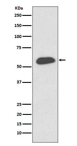
IRF5 Antibody
Rabbit mAb
- SPECIFICATION
- CITATIONS
- PROTOCOLS
- BACKGROUND

Application
| WB, IHC, FC, ICC, IP |
|---|---|
| Primary Accession | Q13568 |
| Reactivity | Rat |
| Clonality | Monoclonal |
| Other Names | Interferon regulatory factor 5; Interferon regulatory factor 5 bone marrow variant; IRF 5; SLEB10; |
| Isotype | Rabbit IgG |
| Host | Rabbit |
| Calculated MW | 56044 Da |
| Dilution | WB 1:500~1:1000 IHC 1:50~1:200 ICC/IF 1:100~1:500 IP 1:100 FC 1:50 |
|---|---|
| Purification | Affinity-chromatography |
| Immunogen | A synthesized peptide derived from human IRF5 |
| Description | Interferon regulatory factors (IRFs) comprise a family of transcription factors that function within the Jak/Stat pathway to regulate interferon (IFN) and IFN-inducible gene expression in response to viral infection. IRFs play an important role in pathogen defense, autoimmunity, lymphocyte development, cell growth, and susceptibility to transformation. |
| Storage Condition and Buffer | Rabbit IgG in phosphate buffered saline , pH 7.4, 150mM NaCl, 0.02% sodium azide and 50% glycerol. Store at +4°C short term. Store at -20°C long term. Avoid freeze / thaw cycle. |
| Name | IRF5 {ECO:0000303|PubMed:11303025, ECO:0000312|HGNC:HGNC:6120} |
|---|---|
| Function | Transcription factor that plays a critical role in innate immunity by activating expression of type I interferon (IFN) IFNA and INFB and inflammatory cytokines downstream of endolysosomal toll-like receptors TLR7, TLR8 and TLR9 (PubMed:11303025, PubMed:15695821, PubMed:22412986, PubMed:25326418, PubMed:32433612). Regulates the transcription of type I IFN genes (IFN-alpha and IFN-beta) and IFN- stimulated genes (ISG) by binding to an interferon-stimulated response element (ISRE) in their promoters (By similarity). Can efficiently activate both the IFN-beta (IFNB) and the IFN-alpha (IFNA) genes and mediate their induction downstream of the TLR-activated, MyD88- dependent pathway (By similarity). Key transcription factor regulating the IFN response during SARS-CoV-2 infection (PubMed:33440148). |
| Cellular Location | Cytoplasm. Nucleus. Note=Shuttles between the nucleus and the cytoplasm: upon activation by the TLR adapter MYD88 and subsequent phosphorylation, translocates to the nucleus |
Research Areas
Citations (0)

Thousands of laboratories across the world have published research that depended on the performance of antibodies from Abcepta to advance their research. Check out links to articles that cite our products in major peer-reviewed journals, organized by research category.
Submit your citation using an Abcepta antibody to
info@abcepta.com, and receive a free "I Love Antibodies" mug.
info@abcepta.com, and receive a free "I Love Antibodies" mug.

Application Protocols
Provided below are standard protocols that you may find useful for product applications.
Abcepta welcomes feedback from its customers.
If you have used an Abcepta product and would like to share how it has performed, please click on the "Submit Review" button and provide the requested information. Our staff will examine and post your review and contact you if needed.
If you have any additional inquiries please email technical services at tech@abcepta.com.
$ 195.00
$ 385.00
Cat# AP90823
Ordering Information
United States
AlbaniaAustraliaAustriaBelgiumBosnia & HerzegovinaBrazilBulgariaCanadaCentral AmericaChinaCroatiaCyprusCzech RepublicDenmarkEstoniaFinlandFranceGermanyGreeceHong KongHungaryIcelandIndiaIndonesiaIrelandIsraelItalyJapanLatviaLithuaniaLuxembourgMacedoniaMalaysiaMaltaMexicoNetherlandsNew ZealandNorwayPakistanPolandPortugalRomaniaSerbiaSingaporeSlovakiaSloveniaSouth AfricaSouth KoreaSpainSwedenSwitzerlandTaiwanTurkeyUnited KingdomUnited StatesVietnamWorldwideOthers
USA Headquarters
(888) 735-7227 / (858) 622-0099 or (858) 875-1900
Other Products
Shipping Information
Domestic orders (in stock items)
Shipped out the same day. Orders placed after 1 PM (PST) will ship out the next business day.
International orders
Contact your local distributors
