


 Foundational characteristics of cancer include proliferation, angiogenesis, migration, evasion of apoptosis, and cellular immortality. Find key markers for these cellular processes and antibodies to detect them.
 The SUMOplot™ Analysis Program predicts and scores sumoylation sites in your protein. SUMOylation is a post-translational modification involved in various cellular processes, such as nuclear-cytosolic transport, transcriptional regulation, apoptosis, protein stability, response to stress, and progression through the cell cycle.
 The Autophagy Receptor Motif Plotter predicts and scores autophagy receptor binding sites in your protein. Identifying proteins connected to this pathway is critical to understanding the role of autophagy in physiological as well as pathological processes such as development, differentiation, neurodegenerative diseases, stress, infection, and cancer.
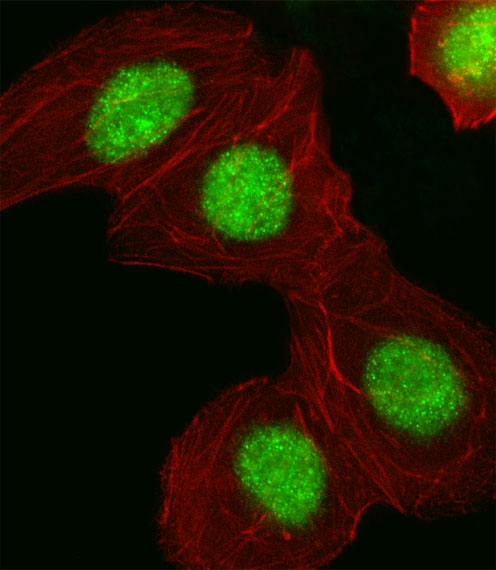
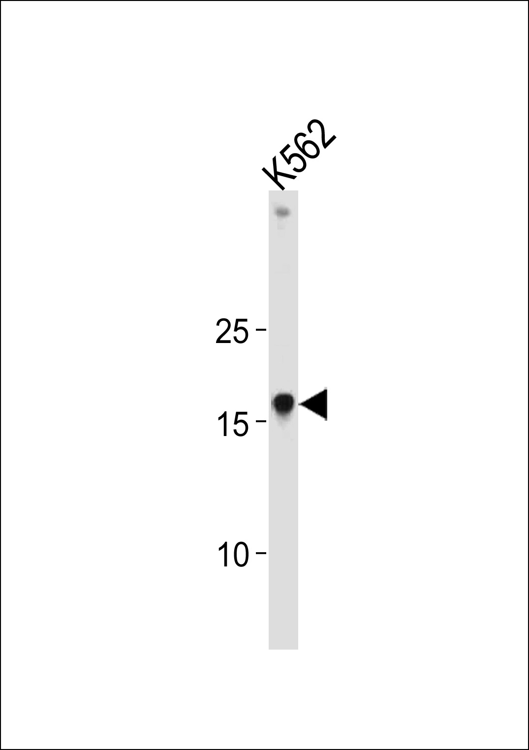
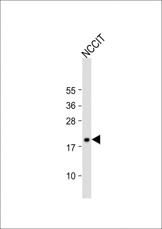
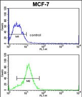
HMGA1 Antibody (C-term)
Purified Rabbit Polyclonal Antibody (Pab)
- SPECIFICATION
- CITATIONS: 1
- PROTOCOLS
- BACKGROUND

Application
| WB, IF, FC, E |
|---|---|
| Primary Accession | P17096 |
| Other Accession | Q8K585, P17095, Q9QXP3 |
| Reactivity | Human |
| Predicted | Hamster, Mouse, Rat |
| Host | Rabbit |
| Clonality | Polyclonal |
| Isotype | Rabbit IgG |
| Calculated MW | 11676 Da |
| Antigen Region | 64-93 aa |
| Gene ID | 3159 |
|---|---|
| Other Names | High mobility group protein HMG-I/HMG-Y, HMG-I(Y), High mobility group AT-hook protein 1, High mobility group protein A1, High mobility group protein R, HMGA1, HMGIY |
| Target/Specificity | This HMGA1 antibody is generated from rabbits immunized with a KLH conjugated synthetic peptide between 64-93 amino acids from the C-terminal region of human HMGA1. |
| Dilution | WB~~1:1000 IF~~1:10~50 FC~~1:10~50 E~~Use at an assay dependent concentration. |
| Format | Purified polyclonal antibody supplied in PBS with 0.09% (W/V) sodium azide. This antibody is prepared by Saturated Ammonium Sulfate (SAS) precipitation followed by dialysis against PBS. |
| Storage | Maintain refrigerated at 2-8°C for up to 2 weeks. For long term storage store at -20°C in small aliquots to prevent freeze-thaw cycles. |
| Precautions | HMGA1 Antibody (C-term) is for research use only and not for use in diagnostic or therapeutic procedures. |
| Name | HMGA1 |
|---|---|
| Synonyms | HMGIY |
| Function | HMG-I/Y bind preferentially to the minor groove of A+T rich regions in double-stranded DNA. It is suggested that these proteins could function in nucleosome phasing and in the 3'-end processing of mRNA transcripts. They are also involved in the transcription regulation of genes containing, or in close proximity to A+T-rich regions. |
| Cellular Location | Nucleus. Chromosome. |

Provided below are standard protocols that you may find useful for product applications.
Background
HMGA1 encodes a non-histone protein involved in many cellular processes, including regulation of inducible gene transcription, integration of retroviruses into chromosomes, and the metastatic progression of cancer cells. The encoded protein preferentially binds to the minor groove of A+T-rich regions in double-stranded DNA. It has little secondary structure in solution but assumes distinct conformations when bound to substrates such as DNA or other proteins. The encoded protein is frequently acetylated and is found in the nucleus.
References
Mu,G., et.al., Hum. Pathol. 41 (4), 493-502 (2010)
Kim,J.J., et.al., J. Hum. Genet. 55 (1), 27-31 (2010)
If you have used an Abcepta product and would like to share how it has performed, please click on the "Submit Review" button and provide the requested information. Our staff will examine and post your review and contact you if needed.
If you have any additional inquiries please email technical services at tech@abcepta.com.
Ordering Information
Other Products
Shipping Information
