


 Foundational characteristics of cancer include proliferation, angiogenesis, migration, evasion of apoptosis, and cellular immortality. Find key markers for these cellular processes and antibodies to detect them.
 The SUMOplot™ Analysis Program predicts and scores sumoylation sites in your protein. SUMOylation is a post-translational modification involved in various cellular processes, such as nuclear-cytosolic transport, transcriptional regulation, apoptosis, protein stability, response to stress, and progression through the cell cycle.
 The Autophagy Receptor Motif Plotter predicts and scores autophagy receptor binding sites in your protein. Identifying proteins connected to this pathway is critical to understanding the role of autophagy in physiological as well as pathological processes such as development, differentiation, neurodegenerative diseases, stress, infection, and cancer.
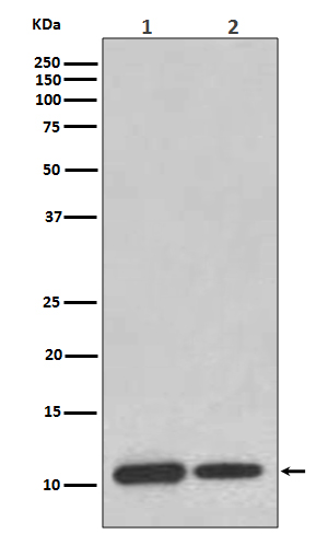
MIF Antibody
Rabbit mAb
- SPECIFICATION
- CITATIONS
- PROTOCOLS
- BACKGROUND

Application
| WB, FC, IP |
|---|---|
| Primary Accession | P14174 |
| Reactivity | Rat |
| Clonality | Monoclonal |
| Other Names | GIF; GLIF; Glycosylation inhibiting factor; L-dopachrome isomerase; MIF protein; MMIF; |
| Isotype | Rabbit IgG |
| Host | Rabbit |
| Calculated MW | 12476 Da |
| Dilution | WB 1:500~1:2000 IP 1:50 FC 1:50 |
|---|---|
| Purification | Affinity-chromatography |
| Immunogen | A synthesized peptide derived from human MIF |
| Description | MIF promotes the systemic inflammatory response by counter-regulating glucocorticoid-mediated inhibition of immune-cell activation and proinflammatory cytokine production. MIF may mediate tissue destruction through the induction of proteinases. |
| Storage Condition and Buffer | Rabbit IgG in phosphate buffered saline , pH 7.4, 150mM NaCl, 0.02% sodium azide and 50% glycerol. Store at +4°C short term. Store at -20°C long term. Avoid freeze / thaw cycle. |
| Name | MIF {ECO:0000303|PubMed:2552447, ECO:0000312|HGNC:HGNC:7097} |
|---|---|
| Function | Pro-inflammatory cytokine involved in the innate immune response to bacterial pathogens (PubMed:15908412, PubMed:17443469, PubMed:23776208). The expression of MIF at sites of inflammation suggests a role as mediator in regulating the function of macrophages in host defense (PubMed:15908412, PubMed:17443469, PubMed:23776208). Counteracts the anti-inflammatory activity of glucocorticoids (PubMed:15908412, PubMed:17443469, PubMed:23776208). Has phenylpyruvate tautomerase and dopachrome tautomerase activity (in vitro), but the physiological substrate is not known (PubMed:11439086, PubMed:17526494). It is not clear whether the tautomerase activity has any physiological relevance, and whether it is important for cytokine activity (PubMed:11439086, PubMed:17526494). |
| Cellular Location | Secreted. Cytoplasm. Note=Does not have a cleavable signal sequence and is secreted via a specialized, non-classical pathway Secreted by macrophages upon stimulation by bacterial lipopolysaccharide (LPS), or by M.tuberculosis antigens |
Research Areas
Citations (0)

Thousands of laboratories across the world have published research that depended on the performance of antibodies from Abcepta to advance their research. Check out links to articles that cite our products in major peer-reviewed journals, organized by research category.
Submit your citation using an Abcepta antibody to
info@abcepta.com, and receive a free "I Love Antibodies" mug.
info@abcepta.com, and receive a free "I Love Antibodies" mug.

Application Protocols
Provided below are standard protocols that you may find useful for product applications.
Abcepta welcomes feedback from its customers.
If you have used an Abcepta product and would like to share how it has performed, please click on the "Submit Review" button and provide the requested information. Our staff will examine and post your review and contact you if needed.
If you have any additional inquiries please email technical services at tech@abcepta.com.
$ 195.00
$ 385.00
Cat# AP91259
Ordering Information
United States
AlbaniaAustraliaAustriaBelgiumBosnia & HerzegovinaBrazilBulgariaCanadaCentral AmericaChinaCroatiaCyprusCzech RepublicDenmarkEstoniaFinlandFranceGermanyGreeceHong KongHungaryIcelandIndiaIndonesiaIrelandIsraelItalyJapanLatviaLithuaniaLuxembourgMacedoniaMalaysiaMaltaMexicoNetherlandsNew ZealandNorwayPakistanPolandPortugalRomaniaSerbiaSingaporeSlovakiaSloveniaSouth AfricaSouth KoreaSpainSwedenSwitzerlandTaiwanTurkeyUnited KingdomUnited StatesVietnamWorldwideOthers
USA Headquarters
(888) 735-7227 / (858) 622-0099 or (858) 875-1900
Other Products
Shipping Information
Domestic orders (in stock items)
Shipped out the same day. Orders placed after 1 PM (PST) will ship out the next business day.
International orders
Contact your local distributors
