


 Foundational characteristics of cancer include proliferation, angiogenesis, migration, evasion of apoptosis, and cellular immortality. Find key markers for these cellular processes and antibodies to detect them.
 The SUMOplot™ Analysis Program predicts and scores sumoylation sites in your protein. SUMOylation is a post-translational modification involved in various cellular processes, such as nuclear-cytosolic transport, transcriptional regulation, apoptosis, protein stability, response to stress, and progression through the cell cycle.
 The Autophagy Receptor Motif Plotter predicts and scores autophagy receptor binding sites in your protein. Identifying proteins connected to this pathway is critical to understanding the role of autophagy in physiological as well as pathological processes such as development, differentiation, neurodegenerative diseases, stress, infection, and cancer.
> home > Products > Primary Antibodies > Antibody Collections > Colon Rectum positive > Desmoglein 2 Antibody
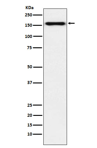
Desmoglein 2 Antibody
Rabbit mAb
- SPECIFICATION
- CITATIONS
- PROTOCOLS
- BACKGROUND

Application
| WB, IHC, FC, ICC |
|---|---|
| Primary Accession | Q14126 |
| Reactivity | Rat |
| Clonality | Monoclonal |
| Other Names | desmoglein-2; DSG2; HDGC; ARVD10; CDHF5; CMD1BB; |
| Isotype | Rabbit IgG |
| Host | Rabbit |
| Calculated MW | 122294 Da |
| Dilution | WB 1:1000~1:5000 IHC 1:50~1:200 ICC/IF 1:50~1:200 FC 1:100 |
|---|---|
| Purification | Affinity-chromatography |
| Immunogen | A synthesized peptide derived from human Desmoglein 2 |
| Description | Component of intercellular desmosome junctions. Involved in the interaction of plaque proteins and intermediate filaments mediating cell-cell adhesion. |
| Storage Condition and Buffer | Rabbit IgG in phosphate buffered saline , pH 7.4, 150mM NaCl, 0.02% sodium azide and 50% glycerol. Store at +4°C short term. Store at -20°C long term. Avoid freeze / thaw cycle. |
| Name | DSG2 |
|---|---|
| Synonyms | CDHF5 |
| Function | A component of desmosome cell-cell junctions which are required for positive regulation of cellular adhesion (PubMed:17559062, PubMed:38395410). Involved in the interaction of plaque proteins and intermediate filaments mediating cell-cell adhesion. Required for proliferation and viability of embryonic stem cells in the blastocyst, thereby crucial for progression of post-implantation embryonic development (By similarity). Maintains pluripotency by regulating epithelial to mesenchymal transition/mesenchymal to epithelial transition (EMT/MET) via interacting with and sequestering CTNNB1 to sites of cell-cell contact, thereby reducing translocation of CTNNB1 to the nucleus and subsequent transcription of CTNNB1/TCF-target genes (PubMed:29910125). Promotes pluripotency and the multi-lineage differentiation potential of hematopoietic stem cells (PubMed:27338829). Plays a role in endothelial cell sprouting and elongation via mediating the junctional-association of cortical actin fibers and CDH5 (PubMed:27338829). Promotes cardiomyocyte cell homeostasis and desmosome junction formation at intercalated disks, as a result plays a role in the maintenance of cardiac conduction and heart chamber integrity (By similarity). Positively regulates pancreatic islet development and maintenance of endothelial cell barrier integrity in the pancreas, therefore involved in the controlled release of insulin from islet cells into the circulation in response to glucose (By similarity). Plays a role in limiting inflammatory infiltration and the apoptotic response to injury in kidney tubular epithelial cells, potentially via its role in maintaining cell-cell adhesion and the epithelial barrier (PubMed:38395410). Acts as a positive modulator of CSK and EGFR activation via sequestering them away from lipid rafts, this is independent of its role in desmosome cell junctions (PubMed:26918609). Also disrupts the localization of CAV1 to lipid rafts resulting in its distribution throughout the cytoplasm (PubMed:26918609). |
| Cellular Location | Cell membrane; Single-pass type I membrane protein. Cell junction, desmosome. Cytoplasm. Note=Localized to intercalated disks in the heart (PubMed:31845994). Localizes to the cytoplasm following cleavage by CASP3 in response to apoptosis (PubMed:17559062) Glycosylation promotes localization to the plasma membrane (PubMed:30885746). |
| Tissue Location | Expressed in undifferentiated pluripotent stem cells, expression decreases during differentiation (at protein level) (PubMed:29910125). Expressed in hematopoietic stem cells and circulating endothelial progenitor cells, expression decreases upon increasing cell lineage commitment (at protein level) (PubMed:27338829). Expressed on common myeloid progenitors, pro- myelocytes, pro-erythrocytes and B-cell linage progenitors (at protein level). Expression in mature cell types in the bone marrow and mature leukocyte populations is absent (PubMed:27338829). Expressed by foreskin fibroblasts, expression peaks during the early stage of differentiation reprogramming (at protein level) (PubMed:29910125) Expressed by endothelial cells in both arterioles and venules in the cervix (at protein level) (PubMed:27338829). Expressed in pancreatic alpha-cells, beta-cells and exocrine tissue (at protein level) (PubMed:36309486). Expressed in cardiomyocytes (at protein level) (PubMed:31845994, PubMed:38375917). Expressed in kidney tubular epithelial cells (PubMed:38395410). |
Research Areas
Citations (0)

Thousands of laboratories across the world have published research that depended on the performance of antibodies from Abcepta to advance their research. Check out links to articles that cite our products in major peer-reviewed journals, organized by research category.
Submit your citation using an Abcepta antibody to
info@abcepta.com, and receive a free "I Love Antibodies" mug.
info@abcepta.com, and receive a free "I Love Antibodies" mug.

Application Protocols
Provided below are standard protocols that you may find useful for product applications.
Abcepta welcomes feedback from its customers.
If you have used an Abcepta product and would like to share how it has performed, please click on the "Submit Review" button and provide the requested information. Our staff will examine and post your review and contact you if needed.
If you have any additional inquiries please email technical services at tech@abcepta.com.
$ 195.00
$ 385.00
Cat# AP91954
Ordering Information
United States
AlbaniaAustraliaAustriaBelgiumBosnia & HerzegovinaBrazilBulgariaCanadaCentral AmericaChinaCroatiaCyprusCzech RepublicDenmarkEstoniaFinlandFranceGermanyGreeceHong KongHungaryIcelandIndiaIndonesiaIrelandIsraelItalyJapanLatviaLithuaniaLuxembourgMacedoniaMalaysiaMaltaMexicoNetherlandsNew ZealandNorwayPakistanPolandPortugalRomaniaSerbiaSingaporeSlovakiaSloveniaSouth AfricaSouth KoreaSpainSwedenSwitzerlandTaiwanTurkeyUnited KingdomUnited StatesVietnamWorldwideOthers
USA Headquarters
(888) 735-7227 / (858) 622-0099 or (858) 875-1900
Other Products
Shipping Information
Domestic orders (in stock items)
Shipped out the same day. Orders placed after 1 PM (PST) will ship out the next business day.
International orders
Contact your local distributors
