


 Foundational characteristics of cancer include proliferation, angiogenesis, migration, evasion of apoptosis, and cellular immortality. Find key markers for these cellular processes and antibodies to detect them.
 The SUMOplot™ Analysis Program predicts and scores sumoylation sites in your protein. SUMOylation is a post-translational modification involved in various cellular processes, such as nuclear-cytosolic transport, transcriptional regulation, apoptosis, protein stability, response to stress, and progression through the cell cycle.
 The Autophagy Receptor Motif Plotter predicts and scores autophagy receptor binding sites in your protein. Identifying proteins connected to this pathway is critical to understanding the role of autophagy in physiological as well as pathological processes such as development, differentiation, neurodegenerative diseases, stress, infection, and cancer.
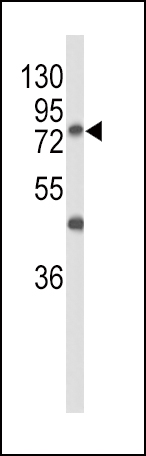
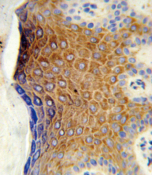
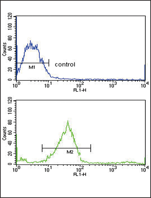
GALNT3 Antibody (Center)
Affinity Purified Rabbit Polyclonal Antibody (Pab)
- SPECIFICATION
- CITATIONS: 1
- PROTOCOLS
- BACKGROUND

Application
| FC, IHC-P, WB, E |
|---|---|
| Primary Accession | Q14435 |
| Reactivity | Human |
| Host | Rabbit |
| Clonality | Polyclonal |
| Isotype | Rabbit IgG |
| Calculated MW | 72610 Da |
| Antigen Region | 454-481 aa |
| Gene ID | 2591 |
|---|---|
| Other Names | Polypeptide N-acetylgalactosaminyltransferase 3, Polypeptide GalNAc transferase 3, GalNAc-T3, pp-GaNTase 3, Protein-UDP acetylgalactosaminyltransferase 3, UDP-GalNAc:polypeptide N-acetylgalactosaminyltransferase 3, GALNT3 |
| Target/Specificity | This GALNT3 antibody is generated from rabbits immunized with a KLH conjugated synthetic peptide between 454-481 amino acids from the Central region of human GALNT3. |
| Dilution | FC~~1:10~50 IHC-P~~1:10~50 WB~~1:1000 E~~Use at an assay dependent concentration. |
| Format | Purified polyclonal antibody supplied in PBS with 0.09% (W/V) sodium azide. This antibody is purified through a protein A column, followed by peptide affinity purification. |
| Storage | Maintain refrigerated at 2-8°C for up to 2 weeks. For long term storage store at -20°C in small aliquots to prevent freeze-thaw cycles. |
| Precautions | GALNT3 Antibody (Center) is for research use only and not for use in diagnostic or therapeutic procedures. |
| Name | GALNT3 |
|---|---|
| Function | Catalyzes the initial reaction in O-linked oligosaccharide biosynthesis, the transfer of an N-acetyl-D-galactosamine residue to a serine or threonine residue on the protein receptor (PubMed:16638743, PubMed:31932717, PubMed:8663203, PubMed:9295285). Has activity toward HIV envelope glycoprotein gp120, EA2, MUC2, MUC1A and MUC5AC (PubMed:8663203, PubMed:9295285). Probably glycosylates fibronectin in vivo (PubMed:9295285). Glycosylates FGF23 (PubMed:16638743, PubMed:31932717). |
| Cellular Location | Golgi apparatus, Golgi stack membrane; Single-pass type II membrane protein. Note=Resides preferentially in the trans and medial parts of the Golgi stack |
| Tissue Location | Expressed in organs that contain secretory epithelial glands. Highly expressed in pancreas, skin, kidney and testis. Weakly expressed in prostate, ovary, intestine and colon. Also expressed in placenta and lung and fetal lung and fetal kidney |

Provided below are standard protocols that you may find useful for product applications.
Background
GALNT3 encodes UDP-GalNAc transferase 3, a member of the GalNAc-transferases family. This family transfers an N-acetyl galactosamine to the hydroxyl group of a serine or threonine residue in the first step of O-linked oligosaccharide biosynthesis. Individual GalNAc-transferases have distinct activities and initiation of O-glycosylation is regulated by a repertoire of GalNAc-transferases. The protein is highly homologous to other family members, however the enzymes have different substrate specificities.
References
Joseph,L., et.al., Skeletal Radiol. 39 (1), 63-68 (2010)
Masi,L.,et.al., J Bone Joint Surg Am 91 (5), 1190-1198 (2009)
Chefetz,I., et.al, Biochim. Biophys. Acta 1792 (1), 61-67 (2009)
If you have used an Abcepta product and would like to share how it has performed, please click on the "Submit Review" button and provide the requested information. Our staff will examine and post your review and contact you if needed.
If you have any additional inquiries please email technical services at tech@abcepta.com.
Ordering Information
Other Products
Shipping Information
