


 Foundational characteristics of cancer include proliferation, angiogenesis, migration, evasion of apoptosis, and cellular immortality. Find key markers for these cellular processes and antibodies to detect them.
 The SUMOplot™ Analysis Program predicts and scores sumoylation sites in your protein. SUMOylation is a post-translational modification involved in various cellular processes, such as nuclear-cytosolic transport, transcriptional regulation, apoptosis, protein stability, response to stress, and progression through the cell cycle.
 The Autophagy Receptor Motif Plotter predicts and scores autophagy receptor binding sites in your protein. Identifying proteins connected to this pathway is critical to understanding the role of autophagy in physiological as well as pathological processes such as development, differentiation, neurodegenerative diseases, stress, infection, and cancer.
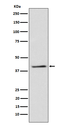
TRIB3 Antibody
Rabbit mAb
- SPECIFICATION
- CITATIONS
- PROTOCOLS
- BACKGROUND

Application
| WB |
|---|---|
| Primary Accession | Q96RU7 |
| Clonality | Monoclonal |
| Other Names | C20orf97; NIPK; SINK; SKIP3; TRB3; Trib3; Tribbles homolog 3; Tribbles pseudokinase 3; Tribbles3; |
| Isotype | Rabbit IgG |
| Host | Rabbit |
| Calculated MW | 39578 Da |
| Dilution | WB 1:500~1:2000 |
|---|---|
| Purification | Affinity-chromatography |
| Immunogen | A synthesized peptide derived from human TRIB3 |
| Description | Disrupts insulin signaling by binding directly to Akt kinases and blocking their activation. May bind directly to and mask the 'Thr-308' phosphorylation site in AKT1. Binds to ATF4 and inhibits its transcriptional activation activity. |
| Storage Condition and Buffer | Rabbit IgG in phosphate buffered saline , pH 7.4, 150mM NaCl, 0.02% sodium azide and 50% glycerol. Store at +4°C short term. Store at -20°C long term. Avoid freeze / thaw cycle. |
| Name | TRIB3 |
|---|---|
| Synonyms | C20orf97, NIPK, SKIP3, TRB3 |
| Function | Inactive protein kinase which acts as a regulator of the integrated stress response (ISR), a process for adaptation to various stress (PubMed:15775988, PubMed:15781252). Inhibits the transcriptional activity of DDIT3/CHOP and is involved in DDIT3/CHOP-dependent cell death during ER stress (PubMed:15775988, PubMed:15781252). May play a role in programmed neuronal cell death but does not appear to affect non-neuronal cells (PubMed:15775988, PubMed:15781252). Acts as a negative feedback regulator of the ATF4-dependent transcription during the ISR: while TRIB3 expression is promoted by ATF4, TRIB3 protein interacts with ATF4 and inhibits ATF4 transcription activity (By similarity). Disrupts insulin signaling by binding directly to Akt kinases and blocking their activation (By similarity). May bind directly to and mask the 'Thr-308' phosphorylation site in AKT1 (By similarity). Interacts with the NF-kappa-B transactivator p65 RELA and inhibits its phosphorylation and thus its transcriptional activation activity (PubMed:12736262). Interacts with MAPK kinases and regulates activation of MAP kinases (PubMed:15299019). Can inhibit APOBEC3A editing of nuclear DNA (PubMed:22977230). |
| Cellular Location | Nucleus. |
| Tissue Location | Highest expression in liver, pancreas, peripheral blood leukocytes and bone marrow. Also highly expressed in a number of primary lung, colon and breast tumors. Expressed in spleen, thymus, and prostate and is undetectable in other examined tissues, including testis, ovary, small intestine, colon, leukocyte, heart, brain, placenta, lung, skeletal muscle, and kidney |
Research Areas
Citations (0)

Thousands of laboratories across the world have published research that depended on the performance of antibodies from Abcepta to advance their research. Check out links to articles that cite our products in major peer-reviewed journals, organized by research category.
Submit your citation using an Abcepta antibody to
info@abcepta.com, and receive a free "I Love Antibodies" mug.
info@abcepta.com, and receive a free "I Love Antibodies" mug.

Application Protocols
Provided below are standard protocols that you may find useful for product applications.
Abcepta welcomes feedback from its customers.
If you have used an Abcepta product and would like to share how it has performed, please click on the "Submit Review" button and provide the requested information. Our staff will examine and post your review and contact you if needed.
If you have any additional inquiries please email technical services at tech@abcepta.com.
$ 195.00
$ 385.00
Cat# AP92159
Ordering Information
United States
AlbaniaAustraliaAustriaBelgiumBosnia & HerzegovinaBrazilBulgariaCanadaCentral AmericaChinaCroatiaCyprusCzech RepublicDenmarkEstoniaFinlandFranceGermanyGreeceHong KongHungaryIcelandIndiaIndonesiaIrelandIsraelItalyJapanLatviaLithuaniaLuxembourgMacedoniaMalaysiaMaltaMexicoNetherlandsNew ZealandNorwayPakistanPolandPortugalRomaniaSerbiaSingaporeSlovakiaSloveniaSouth AfricaSouth KoreaSpainSwedenSwitzerlandTaiwanTurkeyUnited KingdomUnited StatesVietnamWorldwideOthers
USA Headquarters
(888) 735-7227 / (858) 622-0099 or (858) 875-1900
Other Products
Shipping Information
Domestic orders (in stock items)
Shipped out the same day. Orders placed after 1 PM (PST) will ship out the next business day.
International orders
Contact your local distributors
