


 Foundational characteristics of cancer include proliferation, angiogenesis, migration, evasion of apoptosis, and cellular immortality. Find key markers for these cellular processes and antibodies to detect them.
 The SUMOplot™ Analysis Program predicts and scores sumoylation sites in your protein. SUMOylation is a post-translational modification involved in various cellular processes, such as nuclear-cytosolic transport, transcriptional regulation, apoptosis, protein stability, response to stress, and progression through the cell cycle.
 The Autophagy Receptor Motif Plotter predicts and scores autophagy receptor binding sites in your protein. Identifying proteins connected to this pathway is critical to understanding the role of autophagy in physiological as well as pathological processes such as development, differentiation, neurodegenerative diseases, stress, infection, and cancer.
KIR3DL2 Antibody (N-term)
Affinity Purified Rabbit Polyclonal Antibody (Pab)
- SPECIFICATION
- CITATIONS
- PROTOCOLS
- BACKGROUND

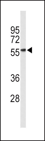
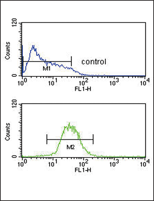
Application
| FC, WB, E |
|---|---|
| Primary Accession | P43630 |
| Reactivity | Human |
| Host | Rabbit |
| Clonality | Polyclonal |
| Isotype | Rabbit IgG |
| Calculated MW | 50230 Da |
| Antigen Region | 46-74 aa |
| Gene ID | 3812 |
|---|---|
| Other Names | Killer cell immunoglobulin-like receptor 3DL2, CD158 antigen-like family member K, MHC class I NK cell receptor, Natural killer-associated transcript 4, NKAT-4, p70 natural killer cell receptor clone CL-5, p70 NK receptor CL-5, CD158k, KIR3DL2, CD158K, NKAT4 |
| Target/Specificity | This KIR3DL2 antibody is generated from rabbits immunized with a KLH conjugated synthetic peptide between 46-74 amino acids from the N-terminal region of human KIR3DL2. |
| Dilution | FC~~1:10~50 WB~~1:1000 E~~Use at an assay dependent concentration. |
| Format | Purified polyclonal antibody supplied in PBS with 0.09% (W/V) sodium azide. This antibody is purified through a protein A column, followed by peptide affinity purification. |
| Storage | Maintain refrigerated at 2-8°C for up to 2 weeks. For long term storage store at -20°C in small aliquots to prevent freeze-thaw cycles. |
| Precautions | KIR3DL2 Antibody (N-term) is for research use only and not for use in diagnostic or therapeutic procedures. |
| Name | KIR3DL2 {ECO:0000303|PubMed:24018270, ECO:0000312|HGNC:HGNC:6339} |
|---|---|
| Function | Receptor on natural killer (NK) cells and T cells for MHC class I molecules (PubMed:24018270, PubMed:28636952). Upon binding of peptide-free HLA-F open conformer, negatively regulates NK and T cell effector functions (PubMed:24018270). Acts as a receptor on astrocytes for HLA-F. Through interaction with HLA-F, may protect motor neurons from astrocyte-induced toxicity (PubMed:26928464). |
| Cellular Location | Cell membrane; Single-pass type I membrane protein |
| Tissue Location | Expressed in astrocytes. |

Thousands of laboratories across the world have published research that depended on the performance of antibodies from Abcepta to advance their research. Check out links to articles that cite our products in major peer-reviewed journals, organized by research category.
info@abcepta.com, and receive a free "I Love Antibodies" mug.

Provided below are standard protocols that you may find useful for product applications.
Background
Killer cell immunoglobulin-like receptors (KIRs) are transmembrane glycoproteins expressed by natural killer cells and subsets of T cells. The KIR genes are polymorphic and highly homologous and they are found in a cluster on chromosome 19q13.4 within the 1 Mb leukocyte receptor complex (LRC). The gene content of the KIR gene cluster varies among haplotypes, although several 'framework' genes are found in all haplotypes (KIR3DL3, KIR3DP1, KIR3DL4, KIR3DL2). The KIR proteins are classified by the number of extracellular immunoglobulin domains (2D or 3D) and by whether they have a long (L) or short (S) cytoplasmic domain. KIR proteins with the long cytoplasmic domain transduce inhibitory signals upon ligand binding via an immune tyrosine-based inhibitory motif (ITIM), while KIR proteins with the short cytoplasmic domain lack the ITIM motif and instead associate with the TYRO protein tyrosine kinase binding protein to transduce activating signals.
References
Pende,D., et.al, J. Exp. Med. 184 (2), 505-518 (1996)
Dohring,C., et.al, Immunogenetics 44 (3), 227-230 (1996)
If you have used an Abcepta product and would like to share how it has performed, please click on the "Submit Review" button and provide the requested information. Our staff will examine and post your review and contact you if needed.
If you have any additional inquiries please email technical services at tech@abcepta.com.
Ordering Information
Other Products
Shipping Information
