


 Foundational characteristics of cancer include proliferation, angiogenesis, migration, evasion of apoptosis, and cellular immortality. Find key markers for these cellular processes and antibodies to detect them.
 The SUMOplot™ Analysis Program predicts and scores sumoylation sites in your protein. SUMOylation is a post-translational modification involved in various cellular processes, such as nuclear-cytosolic transport, transcriptional regulation, apoptosis, protein stability, response to stress, and progression through the cell cycle.
 The Autophagy Receptor Motif Plotter predicts and scores autophagy receptor binding sites in your protein. Identifying proteins connected to this pathway is critical to understanding the role of autophagy in physiological as well as pathological processes such as development, differentiation, neurodegenerative diseases, stress, infection, and cancer.
VAV3 Antibody
Rabbit mAb
- SPECIFICATION
- CITATIONS
- PROTOCOLS
- BACKGROUND

Application
| WB, FC, ICC, IP |
|---|---|
| Primary Accession | Q9UKW4 |
| Clonality | Monoclonal |
| Other Names | RGD1565941; VAV 3; Vav3; VAV3 oncogene; |
| Isotype | Rabbit IgG |
| Host | Rabbit |
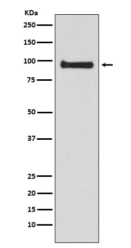
| Calculated MW | 97776 Da |
| Dilution | WB 1:500~1:2000 ICC/IF 1:50~1:200 IP 1:50 FC 1:40. |
|---|---|
| Purification | Affinity-chromatography |
| Immunogen | A synthesized peptide derived from human VAV3 |
| Description | Plays an important role in angiogenesis. Its recruitement by phosphorylated EPHA2 is critical for EFNA1-induced RAC1 GTPase activation and vascular endothelial cell migration and assembly. |
| Storage Condition and Buffer | Rabbit IgG in phosphate buffered saline , pH 7.4, 150mM NaCl, 0.02% sodium azide and 50% glycerol. Store at +4°C short term. Store at -20°C long term. Avoid freeze / thaw cycle. |
| Name | VAV3 |
|---|---|
| Function | Exchange factor for GTP-binding proteins RhoA, RhoG and, to a lesser extent, Rac1. Binds physically to the nucleotide-free states of those GTPases. Plays an important role in angiogenesis. Its recruitment by phosphorylated EPHA2 is critical for EFNA1-induced RAC1 GTPase activation and vascular endothelial cell migration and assembly (By similarity). May be important for integrin-mediated signaling, at least in some cell types. In osteoclasts, along with SYK tyrosine kinase, required for signaling through integrin alpha-v/beta-1 (ITAGV-ITGB1), a crucial event for osteoclast proper cytoskeleton organization and function. This signaling pathway involves RAC1, but not RHO, activation. Necessary for proper wound healing. In the course of wound healing, required for the phagocytotic cup formation preceding macrophage phagocytosis of apoptotic neutrophils. Responsible for integrin beta-2 (ITGB2)-mediated macrophage adhesion and, to a lesser extent, contributes to beta-3 (ITGB3)-mediated adhesion. Does not affect integrin beta-1 (ITGB1)-mediated adhesion (By similarity). |
| Tissue Location | Isoform 1 and isoform 3 are widely expressed; both are expressed at very low levels in skeletal muscle. In keratinocytes, isoform 1 is less abundant than isoform 3. Isoform 3 is detected at very low levels, if any, in adrenal gland, bone marrow, spleen, fetal brain and spinal cord; in these tissues, isoform 1 is readily detectable. |
Research Areas
Citations (0)

Thousands of laboratories across the world have published research that depended on the performance of antibodies from Abcepta to advance their research. Check out links to articles that cite our products in major peer-reviewed journals, organized by research category.
Submit your citation using an Abcepta antibody to
info@abcepta.com, and receive a free "I Love Antibodies" mug.
info@abcepta.com, and receive a free "I Love Antibodies" mug.

Application Protocols
Provided below are standard protocols that you may find useful for product applications.
Abcepta welcomes feedback from its customers.
If you have used an Abcepta product and would like to share how it has performed, please click on the "Submit Review" button and provide the requested information. Our staff will examine and post your review and contact you if needed.
If you have any additional inquiries please email technical services at tech@abcepta.com.
$ 195.00
$ 385.00
Cat# AP92334
Ordering Information
United States
AlbaniaAustraliaAustriaBelgiumBosnia & HerzegovinaBrazilBulgariaCanadaCentral AmericaChinaCroatiaCyprusCzech RepublicDenmarkEstoniaFinlandFranceGermanyGreeceHong KongHungaryIcelandIndiaIndonesiaIrelandIsraelItalyJapanLatviaLithuaniaLuxembourgMacedoniaMalaysiaMaltaMexicoNetherlandsNew ZealandNorwayPakistanPolandPortugalRomaniaSerbiaSingaporeSlovakiaSloveniaSouth AfricaSouth KoreaSpainSwedenSwitzerlandTaiwanTurkeyUnited KingdomUnited StatesVietnamWorldwideOthers
USA Headquarters
(888) 735-7227 / (858) 622-0099 or (858) 875-1900
Other Products
Shipping Information
Domestic orders (in stock items)
Shipped out the same day. Orders placed after 1 PM (PST) will ship out the next business day.
International orders
Contact your local distributors
