


 Foundational characteristics of cancer include proliferation, angiogenesis, migration, evasion of apoptosis, and cellular immortality. Find key markers for these cellular processes and antibodies to detect them.
 The SUMOplot™ Analysis Program predicts and scores sumoylation sites in your protein. SUMOylation is a post-translational modification involved in various cellular processes, such as nuclear-cytosolic transport, transcriptional regulation, apoptosis, protein stability, response to stress, and progression through the cell cycle.
 The Autophagy Receptor Motif Plotter predicts and scores autophagy receptor binding sites in your protein. Identifying proteins connected to this pathway is critical to understanding the role of autophagy in physiological as well as pathological processes such as development, differentiation, neurodegenerative diseases, stress, infection, and cancer.
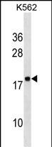
RPLP2 Antibody (N-Term)
Affinity Purified Rabbit Polyclonal Antibody (Pab)
- SPECIFICATION
- CITATIONS
- PROTOCOLS
- BACKGROUND

Application
| WB, E |
|---|---|
| Primary Accession | P05387 |
| Other Accession | P19943, P99027, P42899, NP_000995 |
| Reactivity | Human |
| Predicted | Bovine, Mouse, Rabbit |
| Host | Rabbit |
| Clonality | Polyclonal |
| Isotype | Rabbit IgG |
| Calculated MW | 11665 Da |
| Antigen Region | 13-41 aa |
| Gene ID | 6181 |
|---|---|
| Other Names | 60S acidic ribosomal protein P2, Renal carcinoma antigen NY-REN-44, RPLP2, D11S2243E, RPP2 |
| Target/Specificity | This RPLP2 antibody is generated from rabbits immunized with a KLH conjugated synthetic peptide between 13-41 amino acids from the N-terminal region of human RPLP2. |
| Dilution | WB~~1:1000 E~~Use at an assay dependent concentration. |
| Format | Purified polyclonal antibody supplied in PBS with 0.09% (W/V) sodium azide. This antibody is purified through a protein A column, followed by peptide affinity purification. |
| Storage | Maintain refrigerated at 2-8°C for up to 2 weeks. For long term storage store at -20°C in small aliquots to prevent freeze-thaw cycles. |
| Precautions | RPLP2 Antibody (N-Term) is for research use only and not for use in diagnostic or therapeutic procedures. |
| Name | RPLP2 |
|---|---|
| Synonyms | D11S2243E, RPP2 |
| Function | Plays an important role in the elongation step of protein synthesis. |

Thousands of laboratories across the world have published research that depended on the performance of antibodies from Abcepta to advance their research. Check out links to articles that cite our products in major peer-reviewed journals, organized by research category.
info@abcepta.com, and receive a free "I Love Antibodies" mug.

Provided below are standard protocols that you may find useful for product applications.
Background
RPLP2 consist of a small 40S subunit and a large 60S subunit. Together these subunits are composed of 4 RNA species and approximately 80 structurally distinct proteins. This protein encodes a ribosomal phosphoprotein that is a component of the 60S subunit. The protein, which is a functional equivalent of the E. coli L7/L12 ribosomal protein, belongs to the L12P family of ribosomal proteins. It plays an important role in the elongation step of protein synthesis. Unlike most ribosomal proteins, which are basic, the encoded protein is acidic. Its C-terminal end is nearly identical to the C-terminal ends of the ribosomal phosphoproteins P0 and P1. The P2 protein can interact with P0 and P1 to form a pentameric complex consisting of P1 and P2 dimers, and a P0 monomer. The protein is located in the cytoplasm. As is typical for genes encoding ribosomal proteins, there are multiple processed pseudogenes of this gene dispersed through the genome.
References
Martinez-Azorin,F. FEBS Lett. 582 (20), 3029-3032 (2008) Martinez-Azorin,F. Biochem. J. 413 (3), 527-534 (2008) Sugiyama,N. Mol. Cell Proteomics 6 (6), 1103-1109 (2007)
If you have used an Abcepta product and would like to share how it has performed, please click on the "Submit Review" button and provide the requested information. Our staff will examine and post your review and contact you if needed.
If you have any additional inquiries please email technical services at tech@abcepta.com.
Ordering Information
Other Products
Shipping Information
