


 Foundational characteristics of cancer include proliferation, angiogenesis, migration, evasion of apoptosis, and cellular immortality. Find key markers for these cellular processes and antibodies to detect them.
 The SUMOplot™ Analysis Program predicts and scores sumoylation sites in your protein. SUMOylation is a post-translational modification involved in various cellular processes, such as nuclear-cytosolic transport, transcriptional regulation, apoptosis, protein stability, response to stress, and progression through the cell cycle.
 The Autophagy Receptor Motif Plotter predicts and scores autophagy receptor binding sites in your protein. Identifying proteins connected to this pathway is critical to understanding the role of autophagy in physiological as well as pathological processes such as development, differentiation, neurodegenerative diseases, stress, infection, and cancer.
GPR50 Antibody (Center)
Affinity Purified Rabbit Polyclonal Antibody (Pab)
- SPECIFICATION
- CITATIONS
- PROTOCOLS
- BACKGROUND

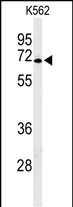
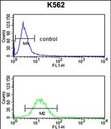
Application
| FC, WB, E |
|---|---|
| Primary Accession | Q13585 |
| Reactivity | Human |
| Host | Rabbit |
| Clonality | Polyclonal |
| Isotype | Rabbit IgG |
| Calculated MW | 67369 Da |
| Antigen Region | 316-344 aa |
| Gene ID | 9248 |
|---|---|
| Other Names | Melatonin-related receptor, G protein-coupled receptor 50, H9, GPR50 |
| Target/Specificity | This GPR50 antibody is generated from rabbits immunized with a KLH conjugated synthetic peptide between 316-344 amino acids from the Central region of human GPR50. |
| Dilution | FC~~1:10~50 WB~~1:1000 E~~Use at an assay dependent concentration. |
| Format | Purified polyclonal antibody supplied in PBS with 0.09% (W/V) sodium azide. This antibody is purified through a protein A column, followed by peptide affinity purification. |
| Storage | Maintain refrigerated at 2-8°C for up to 2 weeks. For long term storage store at -20°C in small aliquots to prevent freeze-thaw cycles. |
| Precautions | GPR50 Antibody (Center) is for research use only and not for use in diagnostic or therapeutic procedures. |
| Name | GPR50 |
|---|---|
| Function | G protein-coupled receptor that plays a role in numerous physiological processes including regulation of energy metabolism, neurite outgrowth or cell migration (PubMed:19699797). Promotes self- renewal and neuronal differentiation of neural progenitor cells through activation of the NOTCH and WNT/beta-catenin signaling pathways (By similarity). Modulates the KAT5-dependent glucocorticoid receptor signaling by modulating KAT5 subcellular compartmentalisation (PubMed:21858214). Also plays a role in the activation TGFBR1 in the absence of TGFBR2 by interfering with FKBP1A binding to TGFBR1, leading to induction of both canonical and non-canonical SMAD signaling pathways resulting in inhibition of proliferation or promotion of migration (PubMed:29572483). |
| Cellular Location | Cell membrane; Multi-pass membrane protein. Postsynaptic density |
| Tissue Location | Hypothalamus and pituitary. |

Thousands of laboratories across the world have published research that depended on the performance of antibodies from Abcepta to advance their research. Check out links to articles that cite our products in major peer-reviewed journals, organized by research category.
info@abcepta.com, and receive a free "I Love Antibodies" mug.

Provided below are standard protocols that you may find useful for product applications.
Background
Melatonin-related receptor expression has been documented from human CNS tissues, specifically in hypothalamus and pituitary, but not in human peripheral tissues. In animals, expression has also been seen in the CNS, and peripheral tissues.
If you have used an Abcepta product and would like to share how it has performed, please click on the "Submit Review" button and provide the requested information. Our staff will examine and post your review and contact you if needed.
If you have any additional inquiries please email technical services at tech@abcepta.com.
Ordering Information
Other Products
Shipping Information
