


 Foundational characteristics of cancer include proliferation, angiogenesis, migration, evasion of apoptosis, and cellular immortality. Find key markers for these cellular processes and antibodies to detect them.
 The SUMOplot™ Analysis Program predicts and scores sumoylation sites in your protein. SUMOylation is a post-translational modification involved in various cellular processes, such as nuclear-cytosolic transport, transcriptional regulation, apoptosis, protein stability, response to stress, and progression through the cell cycle.
 The Autophagy Receptor Motif Plotter predicts and scores autophagy receptor binding sites in your protein. Identifying proteins connected to this pathway is critical to understanding the role of autophagy in physiological as well as pathological processes such as development, differentiation, neurodegenerative diseases, stress, infection, and cancer.
MOTS-C Rabbit Polyclonal Antibody
MOTS-C Rabbit Polyclonal Antibody
- SPECIFICATION
- CITATIONS
- PROTOCOLS
- BACKGROUND

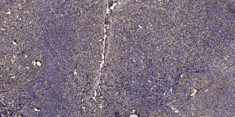
Application
| IHC |
|---|---|
| Primary Accession | A0A0C5B5G6 |
| Reactivity | Rat, Human, Mouse |
| Clonality | Polyclonal |
| Calculated MW | 2175 Da |
| Other Names | Mitochondrial-derived peptide MOTS-c, Mitochondrial open reading frame of the 12S rRNA-c, MT-RNR1 (HGNC:7470) |
|---|---|
| Dilution | IHC~~1:100~500 |
| Storage Conditions | -20℃ |
| Name | MT-RNR1 (HGNC:7470) |
|---|---|
| Function | Regulates insulin sensitivity and metabolic homeostasis (PubMed:25738459, PubMed:33468709). Inhibits the folate cycle, thereby reducing de novo purine biosynthesis which leads to the accumulation of the de novo purine synthesis intermediate 5-aminoimidazole-4- carboxamide (AICAR) and the activation of the metabolic regulator 5'- AMP-activated protein kinase (AMPK) (PubMed:25738459). Protects against age-dependent and diet-induced insulin resistance as well as diet- induced obesity (PubMed:25738459). In response to metabolic stress, translocates to the nucleus where it binds to antioxidant response elements (ARE) present in the promoter regions of a number of genes and plays a role in regulating nuclear gene expression in an NFE2L2- dependent manner and increasing cellular resistance to metabolic stress (PubMed:29983246). Increases mitochondrial respiration and levels of CPT1A and cytokines IL1B, IL6, IL8, IL10 and TNF in senescent cells (PubMed:29886458). Increases activity of the serine/threonine protein kinase complex mTORC2 and reduces activity of the PTEN phosphatase, thus promoting phosphorylation of AKT (PubMed:33554779). This promotes AKT-mediated phosphorylation of transcription factor FOXO1 which reduces FOXO1 activity, leading to reduced levels of MSTN and promotion of skeletal muscle growth (PubMed:33554779). Promotes osteogenic differentiation of bone marrow mesenchymal stem cells via the TGFB/SMAD pathway (PubMed:30468456). Promotes osteoblast proliferation and osteoblast synthesis of type I collagens COL1A1 and COL1A2 via the TGFB/SMAD pathway (PubMed:31081069). |
| Cellular Location | Secreted. Mitochondrion. Nucleus Note=Translocates to the nucleus in response to metabolic stress in an AMPK-dependent manner. |
| Tissue Location | Detected in plasma (at protein level) (PubMed:25738459, PubMed:32182209). Also expressed in skeletal muscle (at protein level) (PubMed:32182209). |
Citations (0)

Thousands of laboratories across the world have published research that depended on the performance of antibodies from Abcepta to advance their research. Check out links to articles that cite our products in major peer-reviewed journals, organized by research category.
Submit your citation using an Abcepta antibody to
info@abcepta.com, and receive a free "I Love Antibodies" mug.
info@abcepta.com, and receive a free "I Love Antibodies" mug.

Application Protocols
Provided below are standard protocols that you may find useful for product applications.
Abcepta welcomes feedback from its customers.
If you have used an Abcepta product and would like to share how it has performed, please click on the "Submit Review" button and provide the requested information. Our staff will examine and post your review and contact you if needed.
If you have any additional inquiries please email technical services at tech@abcepta.com.
$ 385.00
Cat# AP93622
Ordering Information
United States
AlbaniaAustraliaAustriaBelgiumBosnia & HerzegovinaBrazilBulgariaCanadaCentral AmericaChinaCroatiaCyprusCzech RepublicDenmarkEstoniaFinlandFranceGermanyGreeceHong KongHungaryIcelandIndiaIndonesiaIrelandIsraelItalyJapanLatviaLithuaniaLuxembourgMacedoniaMalaysiaMaltaMexicoNetherlandsNew ZealandNorwayPakistanPolandPortugalRomaniaSerbiaSingaporeSlovakiaSloveniaSouth AfricaSouth KoreaSpainSwedenSwitzerlandTaiwanTurkeyUnited KingdomUnited StatesVietnamWorldwideOthers
USA Headquarters
(888) 735-7227 / (858) 622-0099 or (858) 875-1900
Other Products
Shipping Information
Domestic orders (in stock items)
Shipped out the same day. Orders placed after 1 PM (PST) will ship out the next business day.
International orders
Contact your local distributors
