


 Foundational characteristics of cancer include proliferation, angiogenesis, migration, evasion of apoptosis, and cellular immortality. Find key markers for these cellular processes and antibodies to detect them.
 The SUMOplot™ Analysis Program predicts and scores sumoylation sites in your protein. SUMOylation is a post-translational modification involved in various cellular processes, such as nuclear-cytosolic transport, transcriptional regulation, apoptosis, protein stability, response to stress, and progression through the cell cycle.
 The Autophagy Receptor Motif Plotter predicts and scores autophagy receptor binding sites in your protein. Identifying proteins connected to this pathway is critical to understanding the role of autophagy in physiological as well as pathological processes such as development, differentiation, neurodegenerative diseases, stress, infection, and cancer.
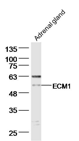
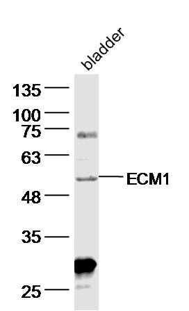
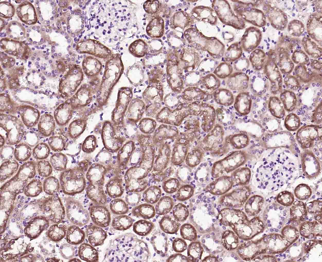
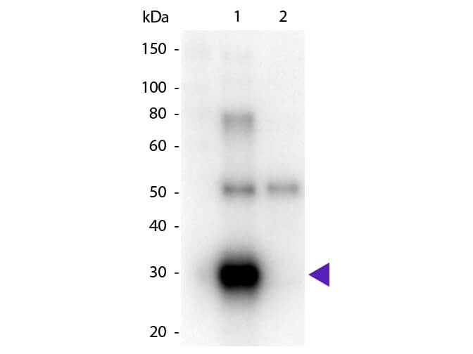
ECM1 Rabbit pAb
ECM1 Rabbit pAb
- SPECIFICATION
- CITATIONS
- PROTOCOLS
- BACKGROUND
Application
| WB, IHC-P, IHC-F, IF |
|---|---|
| Reactivity | Mouse |
| Host | Rabbit |
| Clonality | Polyclonal |
| Calculated MW | 59 KDa |
| Physical State | Liquid |
| Immunogen | KLH conjugated synthetic peptide derived from mouse ECM1 |
| Epitope Specificity | 21-80/567 |
| Isotype | IgG |
| Purity | affinity purified by Protein A |
| Buffer | 0.01M TBS (pH7.4) with 1% BSA, 0.02% Proclin300 and 50% Glycerol. |
| SUBCELLULAR LOCATION | Secreted, extracellular space, extracellular matrix. |
| SUBUNIT | Interacts (via C-terminus) with HSPG2 (via C-terminus). Interacts with EFEMP1/FBLN3 and LAMB3. Interacts with MMP9. |
| DISEASE | Lipoid proteinosis (LiP) [MIM:247100]: Rare autosomal recessive disorder characterized by generalized thickening of skin, mucosae and certain viscera. Classical features include beaded eyelid papules and laryngeal infiltration leading to hoarseness. Histologically, there is widespread deposition of hyaline material and disruption/reduplication of basement membrane. Note=The disease is caused by mutations affecting the gene represented in this entry. |
| Important Note | This product as supplied is intended for research use only, not for use in human, therapeutic or diagnostic applications. |
| Background Descriptions | Extracellular matrix protein 1 (ECM1) This family consists of several eukaryotic extracellular matrix protein 1 (ECM1) sequences. ECM1 has been shown to regulate endochondral bone formation, stimulate the proliferation of endothelial cells and induce angiogenesis. Mutations in the ECM1 gene can cause lipoid proteinosis, a disorder which causes generalised thickening of skin, mucosae and certain viscera. Classical features include beaded eyelid papules and laryngeal infiltration leading to hoarseness. |
| Target/Specificity | Expressed in breast cancer tissues. Little or no expression observed in normal breast tissues. Expressed in skin; wide expression is observed throughout the dermis with minimal expression in the epidermis. |
|---|---|
| Dilution | WB=1:500-2000,IHC-P=1:100-500,IHC-F=1:100-500,IF=1:100-500 |
| Storage | Store at -20 ℃ for one year. Avoid repeated freeze/thaw cycles. When reconstituted in sterile pH 7.4 0.01M PBS or diluent of antibody the antibody is stable for at least two weeks at 2-4 ℃. |

Thousands of laboratories across the world have published research that depended on the performance of antibodies from Abcepta to advance their research. Check out links to articles that cite our products in major peer-reviewed journals, organized by research category.
info@abcepta.com, and receive a free "I Love Antibodies" mug.

Provided below are standard protocols that you may find useful for product applications.
Background
This product as supplied is intended for research use only, not for use in human, therapeutic or diagnostic applications.
If you have used an Abcepta product and would like to share how it has performed, please click on the "Submit Review" button and provide the requested information. Our staff will examine and post your review and contact you if needed.
If you have any additional inquiries please email technical services at tech@abcepta.com.
Ordering Information
Shipping Information
