


 Foundational characteristics of cancer include proliferation, angiogenesis, migration, evasion of apoptosis, and cellular immortality. Find key markers for these cellular processes and antibodies to detect them.
 The SUMOplot™ Analysis Program predicts and scores sumoylation sites in your protein. SUMOylation is a post-translational modification involved in various cellular processes, such as nuclear-cytosolic transport, transcriptional regulation, apoptosis, protein stability, response to stress, and progression through the cell cycle.
 The Autophagy Receptor Motif Plotter predicts and scores autophagy receptor binding sites in your protein. Identifying proteins connected to this pathway is critical to understanding the role of autophagy in physiological as well as pathological processes such as development, differentiation, neurodegenerative diseases, stress, infection, and cancer.
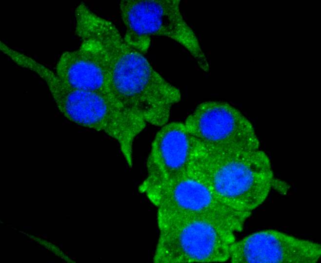
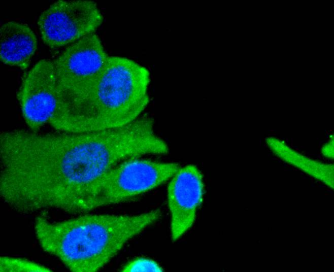
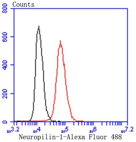
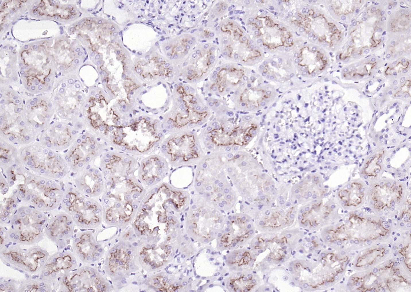
Neuropilin-1 Recombinant Rabbit mAb
Neuropilin-1 Recombinant Rabbit mAb

- SPECIFICATION
- CITATIONS
- PROTOCOLS
- BACKGROUND

Application
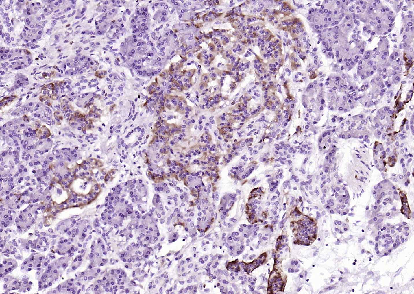
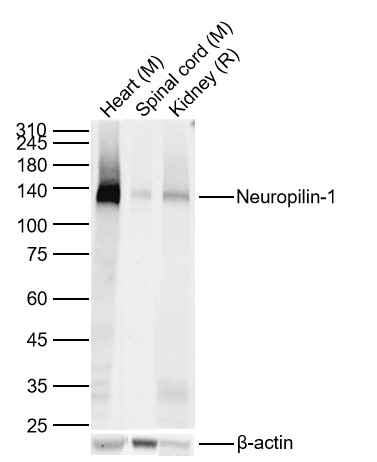
| WB, IHC-P, IHC-F, IF, ICC |
|---|---|
| Primary Accession | Q5T7F3 |
| Reactivity | Human |
| Host | Rabbit |
| Clonality | Recombinant |
| Calculated MW | 103 KDa |
| Physical State | Liquid |
| Immunogen | KLH conjugated synthetic peptide derived from human Neuropilin-1 |
| Purity | affinity purified by Protein A |
| Buffer | 0.01M TBS (pH7.4) with 1% BSA, 0.02% Proclin300 and 50% Glycerol. |
| SUBCELLULAR LOCATION | Cell membrane; Single-pass type I membrane protein. Isoform 2: Secreted. |
| SIMILARITY | Belongs to the neuropilin family. Contains 2 CUB domains. Contains 2 F5/8 type C domains. Contains 1 MAM domain. |
| SUBUNIT | Homodimer, and heterodimer with NRP2. Interacts with FER. Binds PLXNB1. |
| Important Note | This product as supplied is intended for research use only, not for use in human, therapeutic or diagnostic applications. |
| Background Descriptions | This gene encodes one of two neuropilins, which contain specific protein domains which allow them to participate in several different types of signaling pathways that control cell migration. Neuropilins contain a large N-terminal extracellular domain, made up of complement-binding, coagulation factor V/VIII, and meprin domains. These proteins also contains a short membrane-spanning domain and a small cytoplasmic domain. Neuropilins bind many ligands and various types of co-receptors; they affect cell survival, migration, and attraction. Some of the ligands and co-receptors bound by neuropilins are vascular endothelial growth factor (VEGF) and semaphorin family members. Several alternatively spliced transcript variants that encode different protein isoforms have been described for this gene. [provided by RefSeq, Oct 2011] |
| Target/Specificity | The expression of isoforms 1 and 2 does not seem to overlap. Isoform 1 is expressed by the blood vessels of different tissues. In the developing embryo it is found predominantly in the nervous system. In adult tissues, it is highly expressed in heart and placenta; moderately in lung, liver, skeletal muscle, kidney and pancreas; and low in adult brain. Isoform 2 is found in liver hepatocytes, kidney distal and proximal tubules. |
|---|---|
| Dilution | WB=1:500-2000,IHC-P=1:100-500,IHC-F=1:50-200,ICC/IF=1:50,IF=1:100-500,Flow-Cyt=1:50 |
| Storage | Store at -20 ℃ for one year. Avoid repeated freeze/thaw cycles. When reconstituted in sterile pH 7.4 0.01M PBS or diluent of antibody the antibody is stable for at least two weeks at 2-4 ℃. |

Thousands of laboratories across the world have published research that depended on the performance of antibodies from Abcepta to advance their research. Check out links to articles that cite our products in major peer-reviewed journals, organized by research category.
info@abcepta.com, and receive a free "I Love Antibodies" mug.

Provided below are standard protocols that you may find useful for product applications.
Background
This product as supplied is intended for research use only, not for use in human, therapeutic or diagnostic applications.
If you have used an Abcepta product and would like to share how it has performed, please click on the "Submit Review" button and provide the requested information. Our staff will examine and post your review and contact you if needed.
If you have any additional inquiries please email technical services at tech@abcepta.com.
Ordering Information
Other Products
Shipping Information
