


 Foundational characteristics of cancer include proliferation, angiogenesis, migration, evasion of apoptosis, and cellular immortality. Find key markers for these cellular processes and antibodies to detect them.
 The SUMOplot™ Analysis Program predicts and scores sumoylation sites in your protein. SUMOylation is a post-translational modification involved in various cellular processes, such as nuclear-cytosolic transport, transcriptional regulation, apoptosis, protein stability, response to stress, and progression through the cell cycle.
 The Autophagy Receptor Motif Plotter predicts and scores autophagy receptor binding sites in your protein. Identifying proteins connected to this pathway is critical to understanding the role of autophagy in physiological as well as pathological processes such as development, differentiation, neurodegenerative diseases, stress, infection, and cancer.
CCNB1IP1 Antibody (C-term)
Affinity Purified Rabbit Polyclonal Antibody (Pab)
- SPECIFICATION
- CITATIONS
- PROTOCOLS
- BACKGROUND

Application
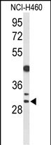
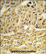
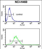
| WB, IHC-P, FC, E |
|---|---|
| Primary Accession | Q9NPC3 |
| Reactivity | Human |
| Host | Rabbit |
| Clonality | Polyclonal |
| Isotype | Rabbit IgG |
| Calculated MW | 31544 Da |
| Antigen Region | 199-228 aa |
| Gene ID | 57820 |
|---|---|
| Other Names | E3 ubiquitin-protein ligase CCNB1IP1, 632-, Cyclin-B1-interacting protein 1, Human enhancer of invasion 10, CCNB1IP1, C14orf18, HEI10 |
| Target/Specificity | This CCNB1IP1 antibody is generated from rabbits immunized with a KLH conjugated synthetic peptide between 199-228 amino acids from the C-terminal region of human CCNB1IP1. |
| Dilution | WB~~1:1000 IHC-P~~1:50~100 FC~~1:10~50 E~~Use at an assay dependent concentration. |
| Format | Purified polyclonal antibody supplied in PBS with 0.09% (W/V) sodium azide. This antibody is purified through a protein A column, followed by peptide affinity purification. |
| Storage | Maintain refrigerated at 2-8°C for up to 2 weeks. For long term storage store at -20°C in small aliquots to prevent freeze-thaw cycles. |
| Precautions | CCNB1IP1 Antibody (C-term) is for research use only and not for use in diagnostic or therapeutic procedures. |
| Name | CCNB1IP1 |
|---|---|
| Synonyms | C14orf18, HEI10 |
| Function | Ubiquitin E3 ligase that acts as a limiting factor for crossing-over during meiosis: required during zygonema to limit the colocalization of RNF212 with MutS-gamma-associated recombination sites and thereby establish early differentiation of crossover and non- crossover sites. Later, it is directed by MutL-gamma to stably accumulate at designated crossover sites. Probably promotes the dissociation of RNF212 and MutS-gamma to allow the progression of recombination and the implementation of the final steps of crossing over (By similarity). Modulates cyclin-B levels and participates in the regulation of cell cycle progression through the G2 phase. Overexpression causes delayed entry into mitosis. |
| Cellular Location | Nucleus. Chromosome. Note=Associates to the synaptonemal complex |
| Tissue Location | Highly expressed in heart. Detected at intermediate levels in liver and kidney, and at low levels in placenta, brain and lung. |

Thousands of laboratories across the world have published research that depended on the performance of antibodies from Abcepta to advance their research. Check out links to articles that cite our products in major peer-reviewed journals, organized by research category.
info@abcepta.com, and receive a free "I Love Antibodies" mug.

Provided below are standard protocols that you may find useful for product applications.
Background
HEI10 is a member of the E3 ubiquitin ligase family and functions in progression of the cell cycle through G(2)/M.
References
Sugiyama, N., et al. Mol. Cell Proteomics 6(6):1103-1109(2007)
Gronholm, M., et al. Oncogene 25(32):4389-4398(2006)
Toby, G.G., et al. Mol. Cell. Biol. 23(6):2109-2122(2003)
If you have used an Abcepta product and would like to share how it has performed, please click on the "Submit Review" button and provide the requested information. Our staff will examine and post your review and contact you if needed.
If you have any additional inquiries please email technical services at tech@abcepta.com.
Ordering Information
Other Products
Shipping Information
