


 Foundational characteristics of cancer include proliferation, angiogenesis, migration, evasion of apoptosis, and cellular immortality. Find key markers for these cellular processes and antibodies to detect them.
 The SUMOplot™ Analysis Program predicts and scores sumoylation sites in your protein. SUMOylation is a post-translational modification involved in various cellular processes, such as nuclear-cytosolic transport, transcriptional regulation, apoptosis, protein stability, response to stress, and progression through the cell cycle.
 The Autophagy Receptor Motif Plotter predicts and scores autophagy receptor binding sites in your protein. Identifying proteins connected to this pathway is critical to understanding the role of autophagy in physiological as well as pathological processes such as development, differentiation, neurodegenerative diseases, stress, infection, and cancer.
LC3B Rabbit pAb
LC3B Rabbit pAb
- SPECIFICATION
- CITATIONS
- PROTOCOLS
- BACKGROUND

Application
| WB |
|---|---|
| Primary Accession | Q9CQV6 |
| Reactivity | Mouse |
| Host | Rabbit |
| Clonality | Polyclonal |
| Calculated MW | 14 KDa |
| Physical State | Liquid |
| Immunogen | KLH conjugated synthetic peptide derived from mouse LC3B |
| Isotype | IgG |
| Purity | affinity purified by Protein A |
| Buffer | 0.01M TBS (pH7.4) with 1% BSA, 0.02% Proclin300 and 50% Glycerol. |
| SUBCELLULAR LOCATION | Cytoplasmic. Endomembrane system; Lipid-anchor. Cytoplasmic vesicle, autophagosome membrane; Lipid-anchor. Note: LC3B binds to the autophagic membranes. |
| SIMILARITY | Belongs to the MAP1 LC3 family. |
| SUBUNIT | 3 different light chains, LC1, LC2 and LC3, can associate with MAP1A and MAP1B proteins. Interacts with SQSTM1. Interacts with TP53INP1 and TP53INP2. |
| Post-translational modifications | The precursor molecule is cleaved by APG4B/ATG4B to form the cytosolic form, LC3-I. This is activated by APG7L/ATG7, transferred to ATG3 and conjugated to phospholipid to form the membrane-bound form, LC3-II. |
| Important Note | This product as supplied is intended for research use only, not for use in human, therapeutic or diagnostic applications. |
| Background Descriptions | A major contributor to cellular homeostasis is the ability of the cell to strike a balance between the formation and degradation/removal of its cellular components. This process of internal cellular turn-over is called autophagy (self-eating), and is facilitated by a pathway of around 16 interacting proteins in the human. LC3, a ubiquitin-like modifier protein, is the human homolog of yeast Apg8 and is involved in the formation of autophagosomal vacuoles, called autophagosomes. LC3 is expressed as 3 splice variants (LC3A, LC3B and LC3C), which exhibit different tissue distributions and are processed into cytosolic and autophagosomal membrane-bound forms, termed LC3-I and LC3-II, respectively. A disruption to the autophagic process is now associated with the progression of several cancers, neurodegenerative disorders and cardiac pathologies, where LC3 is widely employed as a marker for autophagy. |
| Gene ID | 67443 |
|---|---|
| Other Names | Microtubule-associated protein 1 light chain 3 beta, Autophagy-related protein LC3 B, Autophagy-related ubiquitin-like modifier LC3 B, MAP1 light chain 3-like protein 2, Microtubule-associated proteins 1A/1B light chain 3B, MAP1A/MAP1B LC3 B, MAP1A/MAP1B light chain 3 B, Map1lc3b {ECO:0000312|MGI:MGI:1914693}, Map1alc3, Map1lc3 |
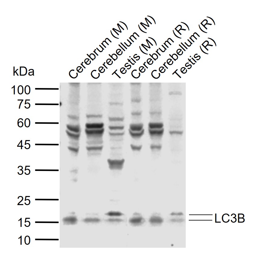
| Target/Specificity | Most abundant in heart, brain, liver, skeletal muscle and testis but absent in thymus and peripheral blood leukocytes. |
| Dilution | WB=1:200-1000 |
| Format | 0.01M TBS(pH7.4), 0.09% (W/V) sodium azide and 50% Glyce |
| Storage | Store at -20 ℃ for one year. Avoid repeated freeze/thaw cycles. When reconstituted in sterile pH 7.4 0.01M PBS or diluent of antibody the antibody is stable for at least two weeks at 2-4 ℃. |
| Name | Map1lc3b {ECO:0000312|MGI:MGI:1914693} |
|---|---|
| Synonyms | Map1alc3, Map1lc3 |
| Function | Ubiquitin-like modifier involved in formation of autophagosomal vacuoles (autophagosomes). Plays a role in mitophagy which contributes to regulate mitochondrial quantity and quality by eliminating the mitochondria to a basal level to fulfill cellular energy requirements and preventing excess ROS production. In response to cellular stress and upon mitochondria fission, binds C-18 ceramides and anchors autophagolysosomes to outer mitochondrial membranes to eliminate damaged mitochondria. While LC3s are involved in elongation of the phagophore membrane, the GABARAP/GATE-16 subfamily is essential for a later stage in autophagosome maturation. Promotes primary ciliogenesis by removing OFD1 from centriolar satellites via the autophagic pathway. Through its interaction with the reticulophagy receptor TEX264, participates in the remodeling of subdomains of the endoplasmic reticulum into autophagosomes upon nutrient stress, which then fuse with lysosomes for endoplasmic reticulum turnover. Upon nutrient stress, directly recruits cofactor JMY to the phagophore membrane surfaces and promotes JMY's actin nucleation activity and autophagosome biogenesis during autophagy. |
| Cellular Location | Cytoplasmic vesicle, autophagosome membrane; Lipid- anchor {ECO:0000250|UniProtKB:Q9GZQ8}. Endomembrane system; Lipid-anchor {ECO:0000250|UniProtKB:Q9GZQ8}. Mitochondrion membrane {ECO:0000250|UniProtKB:Q9GZQ8}; Lipid-anchor {ECO:0000250|UniProtKB:Q9GZQ8}. Cytoplasm, cytoskeleton. Cytoplasmic vesicle {ECO:0000250|UniProtKB:Q9GZQ8}. Note=LC3-II binds to the autophagic membranes. LC3-II localizes with the mitochondrial inner membrane during Parkin-mediated mitophagy (By similarity). Also localizes to discrete punctae along the ciliary axoneme (By similarity) {ECO:0000250|UniProtKB:Q9GZQ8} |

Thousands of laboratories across the world have published research that depended on the performance of antibodies from Abcepta to advance their research. Check out links to articles that cite our products in major peer-reviewed journals, organized by research category.
info@abcepta.com, and receive a free "I Love Antibodies" mug.

Provided below are standard protocols that you may find useful for product applications.
Background
This product as supplied is intended for research use only, not for use in human, therapeutic or diagnostic applications.
If you have used an Abcepta product and would like to share how it has performed, please click on the "Submit Review" button and provide the requested information. Our staff will examine and post your review and contact you if needed.
If you have any additional inquiries please email technical services at tech@abcepta.com.
Ordering Information
Other Products
Shipping Information
