


 Foundational characteristics of cancer include proliferation, angiogenesis, migration, evasion of apoptosis, and cellular immortality. Find key markers for these cellular processes and antibodies to detect them.
 The SUMOplot™ Analysis Program predicts and scores sumoylation sites in your protein. SUMOylation is a post-translational modification involved in various cellular processes, such as nuclear-cytosolic transport, transcriptional regulation, apoptosis, protein stability, response to stress, and progression through the cell cycle.
 The Autophagy Receptor Motif Plotter predicts and scores autophagy receptor binding sites in your protein. Identifying proteins connected to this pathway is critical to understanding the role of autophagy in physiological as well as pathological processes such as development, differentiation, neurodegenerative diseases, stress, infection, and cancer.
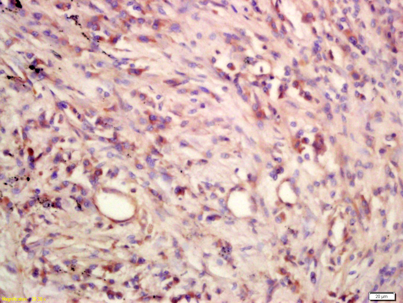
CCL27 Rabbit pAb
CCL27 Rabbit pAb
- SPECIFICATION
- CITATIONS
- PROTOCOLS
- BACKGROUND

Application
| IHC-P, IHC-F, IF |
|---|---|
| Primary Accession | Q9Z1X0 |
| Reactivity | Mouse |
| Host | Rabbit |
| Clonality | Polyclonal |
| Calculated MW | 19 KDa |
| Physical State | Liquid |
| Immunogen | KLH conjugated synthetic peptide derived from mouse CCL27 |
| Epitope Specificity | 25-112/164 |
| Isotype | IgG |
| Purity | affinity purified by Protein A |
| Buffer | 0.01M TBS (pH7.4) with 1% BSA, 0.02% Proclin300 and 50% Glycerol. |
| SUBCELLULAR LOCATION | Secreted. |
| SIMILARITY | Belongs to the intercrine beta (chemokine CC) family. |
| SUBUNIT | Monomer, dimer, and tetramer. Heparin avidly promotes oligomerization. |
| Important Note | This product as supplied is intended for research use only, not for use in human, therapeutic or diagnostic applications. |
| Background Descriptions | This gene is one of several CC cytokine genes clustered on the p-arm of chromosome 9. Cytokines are a family of secreted proteins involved in immunoregulatory and inflammatory processes. The CC cytokines are proteins characterized by two adjacent cysteines. The protein encoded by this gene is chemotactic for skin-associated memory T lymphocytes. This cytokine may also play a role in mediating homing of lymphocytes to cutaneous sites. It specifically binds to chemokine receptor 10 (CCR10). Studies of a similar murine protein indicate that these protein-receptor interactions have a pivotal role in T cell-mediated skin inflammation. [provided by RefSeq]. |
| Gene ID | 20301 |
|---|---|
| Other Names | C-C motif chemokine 27, CC chemokine ILC, Cutaneous T-cell-attracting chemokine, CTACK, ESkine, IL-11 R-alpha-locus chemokine, ALP, mILC, Skinkine, Small-inducible cytokine A27, Ccl27, Ilc, Scya27 |
| Target/Specificity | Testis, thymus, placenta, ovary and skin. |
| Dilution | IHC-P=1:100-500,IHC-F=1:100-500,IF=1:100-500 |
| Storage | Store at -20 ℃ for one year. Avoid repeated freeze/thaw cycles. When reconstituted in sterile pH 7.4 0.01M PBS or diluent of antibody the antibody is stable for at least two weeks at 2-4 ℃. |
| Name | Ccl27 |
|---|---|
| Synonyms | Ilc, Scya27 |
| Function | Chemotactic factor that attracts skin-associated memory T- lymphocytes. May play a role in mediating homing of lymphocytes to cutaneous sites. May play a role in cell migration during embryogenesis. Nuclear forms may facilitate cellular migration by inducing cytoskeletal relaxation. Binds to CCR10. |
| Cellular Location | [Isoform 1]: Secreted. Nucleus Note=May also be nuclear when following receptor (CCR10)-mediated internalization. |
| Tissue Location | Isoform 1 is predominantly expressed in placenta and weakly in skin. Isoform 2 is predominantly expressed in testes and brain, weakly in kidney and liver and even lower in heart and muscle Low expression of both isoforms in other tissues |

Thousands of laboratories across the world have published research that depended on the performance of antibodies from Abcepta to advance their research. Check out links to articles that cite our products in major peer-reviewed journals, organized by research category.
info@abcepta.com, and receive a free "I Love Antibodies" mug.

Provided below are standard protocols that you may find useful for product applications.
Background
This product as supplied is intended for research use only, not for use in human, therapeutic or diagnostic applications.
If you have used an Abcepta product and would like to share how it has performed, please click on the "Submit Review" button and provide the requested information. Our staff will examine and post your review and contact you if needed.
If you have any additional inquiries please email technical services at tech@abcepta.com.
Ordering Information
Other Products
Shipping Information
