


 Foundational characteristics of cancer include proliferation, angiogenesis, migration, evasion of apoptosis, and cellular immortality. Find key markers for these cellular processes and antibodies to detect them.
 The SUMOplot™ Analysis Program predicts and scores sumoylation sites in your protein. SUMOylation is a post-translational modification involved in various cellular processes, such as nuclear-cytosolic transport, transcriptional regulation, apoptosis, protein stability, response to stress, and progression through the cell cycle.
 The Autophagy Receptor Motif Plotter predicts and scores autophagy receptor binding sites in your protein. Identifying proteins connected to this pathway is critical to understanding the role of autophagy in physiological as well as pathological processes such as development, differentiation, neurodegenerative diseases, stress, infection, and cancer.
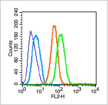
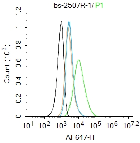
CD44 Rabbit pAb
CD44 Rabbit pAb
- SPECIFICATION
- CITATIONS
- PROTOCOLS
- BACKGROUND

| Primary Accession | P26051 |
|---|---|
| Reactivity | Rat |
| Host | Rabbit |
| Clonality | Polyclonal |
| Calculated MW | 53 KDa |
| Physical State | Liquid |
| Immunogen | KLH conjugated synthetic peptide derived from rat CD44v6 |
| Epitope Specificity | 325-429/429 |
| Isotype | IgG |
| Purity | affinity purified by Protein A |
| Buffer | 0.01M TBS (pH7.4) with 1% BSA, 0.02% Proclin300 and 50% Glycerol. |
| SUBCELLULAR LOCATION | Membrane; Single-pass type I membrane protein. Note=Colocalizes with actin in membrane protrusions at wounding edges. |
| SIMILARITY | Contains 1 Link domain. |
| SUBUNIT | Interacts with PKN2 (By similarity). Interacts with HA, as well as other glycosaminoglycans, collagen, laminin, and fibronectin via its N-terminal segment. Interacts with ANK, the ERM proteins (VIL2, RDX and MSN), and NF2 via its C-terminal segment. |
| Post-translational modifications | Proteolytically cleaved in the extracellular matrix by specific proteinases (possibly MMPs) in several cell lines and tumors. N- and O-glycosylated. O-glycosylation contains more-or-less-sulfated chondroitin sulfate glycans, whose number may affect the accessibility of specific proteinases to their cleavage site(s). It is uncertain if O-glycosylation occurs on Thr-637 or Thr-638. Phosphorylated; activation of PKC results in the dephosphorylation of Ser-706 (constitutive phosphorylation site), and the phosphorylation of Ser-672. |
| Important Note | This product as supplied is intended for research use only, not for use in human, therapeutic or diagnostic applications. |
| Background Descriptions | The protein encoded by this gene is a cell-surface glycoprotein involved in cell-cell interactions, cell adhesion and migration. It is a receptor for hyaluronic acid (HA) and can also interact with other ligands, such as osteopontin, collagens, and matrix metalloproteinases (MMPs). This protein participates in a wide variety of cellular functions including lymphocyte activation, recirculation and homing, hematopoiesis, and tumor metastasis. Transcripts for this gene undergo complex alternative splicing that results in many functionally distinct isoforms, however, the full length nature of some of these variants has not been determined. Alternative splicing is the basis for the structural and functional diversity of this protein, and may be related to tumor metastasis. [provided by RefSeq, Jul 2008]. |
| Gene ID | 25406 |
|---|---|
| Other Names | CD44 antigen, Extracellular matrix receptor III, ECMR-III, GP90 lymphocyte homing/adhesion receptor, HUTCH-I, Hermes antigen, Hyaluronate receptor, Phagocytic glycoprotein 1, PGP-1, Phagocytic glycoprotein I, PGP-I, CD44, Cd44 |
| Target/Specificity | Isoform 10 (epithelial isoform) is expressed by cells of epithelium and highly expressed by carcinomas. Expression is repressed in neuroblastoma cells. |
| Dilution | Flow-Cyt=1 µg/Test |
| Storage | Store at -20 ℃ for one year. Avoid repeated freeze/thaw cycles. When reconstituted in sterile pH 7.4 0.01M PBS or diluent of antibody the antibody is stable for at least two weeks at 2-4 ℃. |
| Name | Cd44 |
|---|---|
| Function | Cell-surface receptor that plays a role in cell-cell interactions, cell adhesion and migration, helping them to sense and respond to changes in the tissue microenvironment. Participates thereby in a wide variety of cellular functions including the activation, recirculation and homing of T-lymphocytes, hematopoiesis, inflammation and response to bacterial infection. Engages, through its ectodomain, extracellular matrix components such as hyaluronan/HA, collagen, growth factors, cytokines or proteases and serves as a platform for signal transduction by assembling, via its cytoplasmic domain, protein complexes containing receptor kinases and membrane proteases. Such effectors include PKN2, the RhoGTPases RAC1 and RHOA, Rho-kinases and phospholipase C that coordinate signaling pathways promoting calcium mobilization and actin-mediated cytoskeleton reorganization essential for cell migration and adhesion. |
| Cellular Location | Cell membrane {ECO:0000250|UniProtKB:P15379}; Single-pass type I membrane protein {ECO:0000250|UniProtKB:P15379} Cell projection, microvillus {ECO:0000250|UniProtKB:P15379}. Secreted {ECO:0000250|UniProtKB:P16070}. Note=Colocalizes with actin in membrane protrusions at wounding edges. Co-localizes with RDX, EZR and MSN in microvilli. {ECO:0000250|UniProtKB:P15379} |

Thousands of laboratories across the world have published research that depended on the performance of antibodies from Abcepta to advance their research. Check out links to articles that cite our products in major peer-reviewed journals, organized by research category.
info@abcepta.com, and receive a free "I Love Antibodies" mug.

Provided below are standard protocols that you may find useful for product applications.
Background
This product as supplied is intended for research use only, not for use in human, therapeutic or diagnostic applications.
If you have used an Abcepta product and would like to share how it has performed, please click on the "Submit Review" button and provide the requested information. Our staff will examine and post your review and contact you if needed.
If you have any additional inquiries please email technical services at tech@abcepta.com.
Ordering Information
Other Products
Shipping Information
