


 Foundational characteristics of cancer include proliferation, angiogenesis, migration, evasion of apoptosis, and cellular immortality. Find key markers for these cellular processes and antibodies to detect them.
 The SUMOplot™ Analysis Program predicts and scores sumoylation sites in your protein. SUMOylation is a post-translational modification involved in various cellular processes, such as nuclear-cytosolic transport, transcriptional regulation, apoptosis, protein stability, response to stress, and progression through the cell cycle.
 The Autophagy Receptor Motif Plotter predicts and scores autophagy receptor binding sites in your protein. Identifying proteins connected to this pathway is critical to understanding the role of autophagy in physiological as well as pathological processes such as development, differentiation, neurodegenerative diseases, stress, infection, and cancer.
ACP1 Antibody (N-term)
Affinity Purified Rabbit Polyclonal Antibody (Pab)
- SPECIFICATION
- CITATIONS
- PROTOCOLS
- BACKGROUND

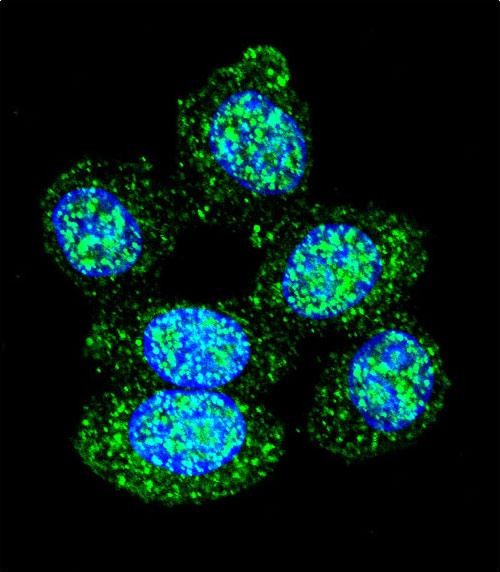
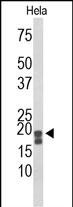
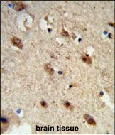
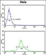
Application
| IF, FC, IHC-P, WB, E |
|---|---|
| Primary Accession | P24666 |
| Reactivity | Human |
| Host | Rabbit |
| Clonality | Polyclonal |
| Isotype | Rabbit IgG |
| Calculated MW | 18042 Da |
| Antigen Region | 33-61 aa |
| Gene ID | 52 |
|---|---|
| Other Names | Low molecular weight phosphotyrosine protein phosphatase, LMW-PTP, LMW-PTPase, Adipocyte acid phosphatase, Low molecular weight cytosolic acid phosphatase, Red cell acid phosphatase 1, ACP1 |
| Target/Specificity | This ACP1 antibody is generated from rabbits immunized with a KLH conjugated synthetic peptide between 33-61 amino acids from the N-terminal region of human ACP1. |
| Dilution | IF~~1:10~50 FC~~1:10~50 IHC-P~~1:50~100 WB~~1:1000 E~~Use at an assay dependent concentration. |
| Format | Purified polyclonal antibody supplied in PBS with 0.09% (W/V) sodium azide. This antibody is purified through a protein A column, followed by peptide affinity purification. |
| Storage | Maintain refrigerated at 2-8°C for up to 2 weeks. For long term storage store at -20°C in small aliquots to prevent freeze-thaw cycles. |
| Precautions | ACP1 Antibody (N-term) is for research use only and not for use in diagnostic or therapeutic procedures. |
| Name | ACP1 (HGNC:122) |
|---|---|
| Function | Acts on tyrosine phosphorylated proteins, low-MW aryl phosphates and natural and synthetic acyl phosphates with differences in substrate specificity between isoform 1 and isoform 2. |
| Cellular Location | Cytoplasm. |
| Tissue Location | [Isoform 2]: Expressed in T-lymphocytes. |

Thousands of laboratories across the world have published research that depended on the performance of antibodies from Abcepta to advance their research. Check out links to articles that cite our products in major peer-reviewed journals, organized by research category.
info@abcepta.com, and receive a free "I Love Antibodies" mug.

Provided below are standard protocols that you may find useful for product applications.
Background
ACP1 belongs to the phosphotyrosine protein phosphatase family of proteins. It functions as an acid phosphatase and a protein tyrosine phosphatase by hydrolyzing protein tyrosine phosphate to protein tyrosine and orthophosphate. This enzyme also hydrolyzes orthophosphoric monoesters to alcohol and orthophosphate. This gene is genetically polymorphic, and three common alleles segregating at the corresponding locus give rise to six phenotypes. Each allele appears to encode at least two electrophoretically different isozymes, Bf and Bs, which are produced in allele-specific ratios.
References
Saccucci, P., et al. Med. Sci. Monit. 15 (10), CR511-CR517 (2009) : Shu, Y.H., et al. J. Clin. Endocrinol. Metab. 94(10):4094-4102(2009) Apelt, N., et al. Metab. Clin. Exp. 58(10):1415-1423(2009) Banci, M., et al. Cardiology 113(4):236-242(2009) Rousseff, R.T., et al. Neuropediatrics 39(6):354-356(2008)
If you have used an Abcepta product and would like to share how it has performed, please click on the "Submit Review" button and provide the requested information. Our staff will examine and post your review and contact you if needed.
If you have any additional inquiries please email technical services at tech@abcepta.com.
Ordering Information
Other Products
Shipping Information
