


 Foundational characteristics of cancer include proliferation, angiogenesis, migration, evasion of apoptosis, and cellular immortality. Find key markers for these cellular processes and antibodies to detect them.
 The SUMOplot™ Analysis Program predicts and scores sumoylation sites in your protein. SUMOylation is a post-translational modification involved in various cellular processes, such as nuclear-cytosolic transport, transcriptional regulation, apoptosis, protein stability, response to stress, and progression through the cell cycle.
 The Autophagy Receptor Motif Plotter predicts and scores autophagy receptor binding sites in your protein. Identifying proteins connected to this pathway is critical to understanding the role of autophagy in physiological as well as pathological processes such as development, differentiation, neurodegenerative diseases, stress, infection, and cancer.
CPLX1 Antibody (Center)
Affinity Purified Rabbit Polyclonal Antibody (Pab)
- SPECIFICATION
- CITATIONS
- PROTOCOLS
- BACKGROUND

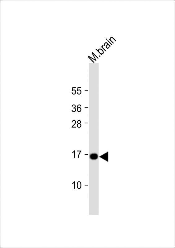
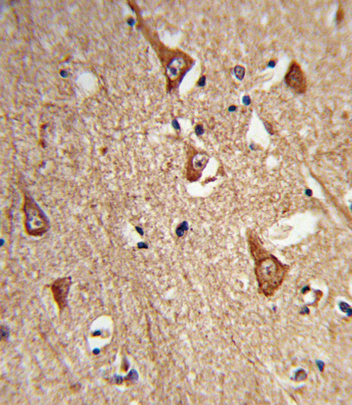
Application
| IHC-P, WB, E |
|---|---|
| Primary Accession | O14810 |
| Other Accession | P63041, P63040, Q4R4N1, Q0IIL7 |
| Reactivity | Mouse |
| Predicted | Bovine, Monkey, Rat |
| Host | Rabbit |
| Clonality | Polyclonal |
| Isotype | Rabbit IgG |
| Calculated MW | 15030 Da |
| Antigen Region | 33-60 aa |
| Gene ID | 10815 |
|---|---|
| Other Names | Complexin-1, Complexin I, CPX I, Synaphin-2, CPLX1 |
| Target/Specificity | This CPLX1 antibody is generated from rabbits immunized with a KLH conjugated synthetic peptide between 33-60 amino acids from the Central region of human CPLX1. |
| Dilution | IHC-P~~1:10~50 WB~~1:1000 E~~Use at an assay dependent concentration. |
| Format | Purified polyclonal antibody supplied in PBS with 0.09% (W/V) sodium azide. This antibody is purified through a protein A column, followed by peptide affinity purification. |
| Storage | Maintain refrigerated at 2-8°C for up to 2 weeks. For long term storage store at -20°C in small aliquots to prevent freeze-thaw cycles. |
| Precautions | CPLX1 Antibody (Center) is for research use only and not for use in diagnostic or therapeutic procedures. |
| Name | CPLX1 |
|---|---|
| Function | Positively regulates a late step in exocytosis of various cytoplasmic vesicles, such as synaptic vesicles and other secretory vesicles (PubMed:21785414). Organizes the SNAREs into a cross-linked zigzag topology that, when interposed between the vesicle and plasma membranes, is incompatible with fusion, thereby preventing SNAREs from releasing neurotransmitters until an action potential arrives at the synapse (PubMed:21785414). Also involved in glucose-induced secretion of insulin by pancreatic beta-cells. Essential for motor behavior. |
| Cellular Location | Cytoplasm, cytosol {ECO:0000250|UniProtKB:P63040}. Perikaryon {ECO:0000250|UniProtKB:P63040}. Presynapse {ECO:0000250|UniProtKB:P63040}. Note=Enriched at synaptic-releasing sites in mature neurons. {ECO:0000250|UniProtKB:P63040} |
| Tissue Location | Nervous system. In hippocampus and cerebellum, expressed mainly by inhibitory neurons. Overexpressed in substantia nigra from patients with Parkinson disease |

Thousands of laboratories across the world have published research that depended on the performance of antibodies from Abcepta to advance their research. Check out links to articles that cite our products in major peer-reviewed journals, organized by research category.
info@abcepta.com, and receive a free "I Love Antibodies" mug.

Provided below are standard protocols that you may find useful for product applications.
Background
CPLX1 encoded by the complexin/synaphin gene family are cytosolic proteins that function in synaptic vesicle exocytosis. These proteins bind syntaxin, part of the SNAP receptor. The protein product of this gene binds to the SNAP receptor complex and disrupts it, allowing transmitter release.
References
Salimi, K., et al. Synapse 62(4):273-282(2008)
Giraudo, C.G., et al. Science 313(5787):676-680(2006)
Kishi, T., et al. Schizophr. Res. 82 (2-3), 185-189 (2006) :
Basso, M., et al. Proteomics 4(12):3943-3952(2004)
Chen, X., et al. Neuron 33(3):397-409(2002)
If you have used an Abcepta product and would like to share how it has performed, please click on the "Submit Review" button and provide the requested information. Our staff will examine and post your review and contact you if needed.
If you have any additional inquiries please email technical services at tech@abcepta.com.
Ordering Information
Other Products
Shipping Information
