


 Foundational characteristics of cancer include proliferation, angiogenesis, migration, evasion of apoptosis, and cellular immortality. Find key markers for these cellular processes and antibodies to detect them.
 The SUMOplot™ Analysis Program predicts and scores sumoylation sites in your protein. SUMOylation is a post-translational modification involved in various cellular processes, such as nuclear-cytosolic transport, transcriptional regulation, apoptosis, protein stability, response to stress, and progression through the cell cycle.
 The Autophagy Receptor Motif Plotter predicts and scores autophagy receptor binding sites in your protein. Identifying proteins connected to this pathway is critical to understanding the role of autophagy in physiological as well as pathological processes such as development, differentiation, neurodegenerative diseases, stress, infection, and cancer.
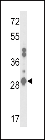
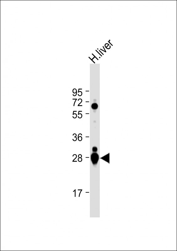
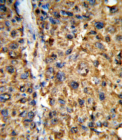
GSTA2 Antibody (N-term)
Affinity Purified Rabbit Polyclonal Antibody (Pab)
- SPECIFICATION
- CITATIONS: 2
- PROTOCOLS
- BACKGROUND

Application
| IHC-P, WB, E |
|---|---|
| Primary Accession | P09210 |
| Other Accession | P08263, Q16772, Q7RTV2 |
| Reactivity | Human |
| Host | Rabbit |
| Clonality | Polyclonal |
| Isotype | Rabbit IgG |
| Calculated MW | 25664 Da |
| Antigen Region | 1-30 aa |
| Gene ID | 2939 |
|---|---|
| Other Names | Glutathione S-transferase A2, GST HA subunit 2, GST class-alpha member 2, GST-gamma, GSTA2-2, GTH2, GSTA2, GST2 |
| Target/Specificity | This GSTA2 antibody is generated from rabbits immunized with a KLH conjugated synthetic peptide between 1-30 amino acids from the N-terminal region of human GSTA2. |
| Dilution | IHC-P~~1:10~50 WB~~1:1000 E~~Use at an assay dependent concentration. |
| Format | Purified polyclonal antibody supplied in PBS with 0.09% (W/V) sodium azide. This antibody is purified through a protein A column, followed by peptide affinity purification. |
| Storage | Maintain refrigerated at 2-8°C for up to 2 weeks. For long term storage store at -20°C in small aliquots to prevent freeze-thaw cycles. |
| Precautions | GSTA2 Antibody (N-term) is for research use only and not for use in diagnostic or therapeutic procedures. |
| Name | GSTA2 |
|---|---|
| Synonyms | GST2 |
| Function | Catalyzes the conjugation of glutathione to a large variety of electrophilic compounds. |
| Cellular Location | Cytoplasm. |
| Tissue Location | Liver.. |

Provided below are standard protocols that you may find useful for product applications.
Background
Cytosolic and membrane-bound forms of glutathione S-transferase are encoded by two distinct supergene families. These enzymes function in the detoxification of electrophilic compounds, including carcinogens, therapeutic drugs, environmental toxins and products of oxidative stress, by conjugation with glutathione. The genes encoding these enzymes are known to be highly polymorphic. These genetic variations can change an individual's susceptibility to carcinogens and toxins as well as affect the toxicity and efficacy of some drugs. At present, eight distinct classes of the soluble cytoplasmic mammalian glutathione S-transferases have been identified: alpha, kappa, mu, omega, pi, sigma, theta and zeta. This gene encodes a glutathione S-tranferase belonging to the alpha class. The alpha class genes, located in a cluster mapped to chromosome 6, are the most abundantly expressed glutathione S-transferases in liver. In addition to metabolizing bilirubin and certain anti-cancer drugs in the liver, the alpha class of these enzymes exhibit glutathione peroxidase activity thereby protecting the cells from reactive oxygen species and the products of peroxidation.
References
Tars, K., et al. J. Mol. Biol. 397(1):332-340(2010) Moyer, A.M., et al. Cancer Epidemiol. Biomarkers Prev. 19(3):811-821(2010) Gemignani, F., et al. Mutat. Res. 671 (1-2), 76-83 (2009) Rohrdanz, E., et al. Arch. Biochem. Biophys. 298(2):747-752(1992) Bogaards, J.J., et al. Biochem. J. 286 (PT 2), 383-388 (1992) Klone, A., et al. Biochem. J. 285 (PT 3), 925-928 (1992)
If you have used an Abcepta product and would like to share how it has performed, please click on the "Submit Review" button and provide the requested information. Our staff will examine and post your review and contact you if needed.
If you have any additional inquiries please email technical services at tech@abcepta.com.
Ordering Information
Shipping Information
